Download presentation
Presentation is loading. Please wait.

1
GY case presentation ( 서 0 나 F/12) 가천의과대학 길 병원 산부인과 Sonographer : 안 소 영
 가천의과대학 길 병원 산부인과 Sonographer : 안 소 영")
2
초음파 통계 OBGY Dopple r T- SO NO amnio3DBPPTOTAL 07/4/9~0 7/4/14 10219919100312 07/4/16~ 07/4/2 1 8520105101293 17214001-19

3
- C.C. : left paraumbilical & lower abdominal pain - P.I. : 위 상기 주 증상으로 abdo.sono, CT 검사에 서 Lt. kidney agenesis, hematocolpos 로 PD 과에서 consult 의뢰되어 GO 외래로 오심.

4
OB/HX : 0-0-0-0 P/HX: (-) F/HX: 친부 : 당뇨 친조모 : 대장암으로 사망 Menstrual history LMP: 2007. 2 월 말 menarche: 2006.9 3~4days Hb 14.4 EKG: normal Chest PA : normal

5
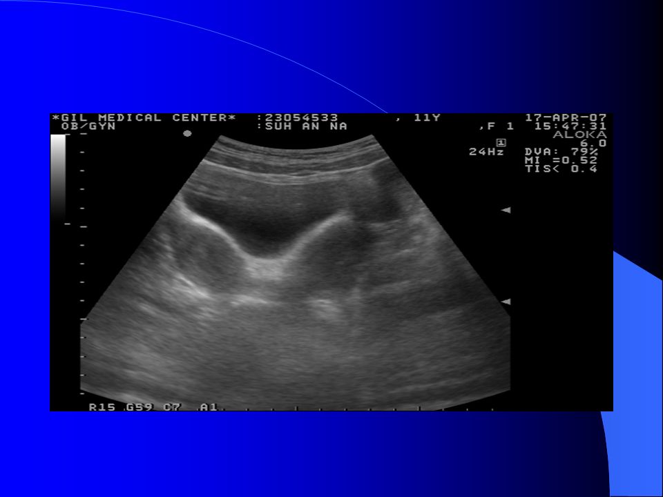
Abdo. Sono 2/23

6
Abdo. sono-IMP No visualization of left kidney. ---> R/O Unilateral renal agenesis, left. Well defined cavity between bladder and rectum. ---> R/O Hydrocolpos. Suspicious bicornuate uterus. REC) APCT.
 APCT..")
7
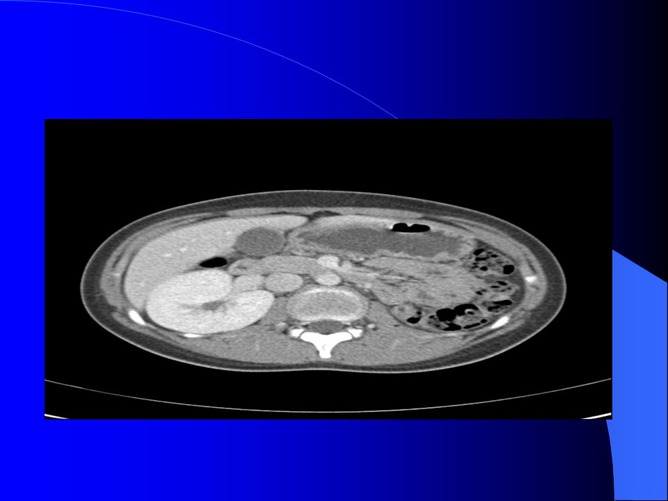
07/3/3

10
CT - IMP 1. Absent left kidney with compensatory hypertrophy of the right kidney. 2. Two uterine horns (Uterine didelphys). Obstruction at left side below cervical level with hematometrocolpos. No evidence of obstruction in the right side uterine horn. -----> Mullerian duct dupilcation anomaly with unilateral kidney. 3. About 5.7 x 4.4 cm sized multicystic lesion in left adnexa. ----> R/O left ovarian cystic lesion. R/O hydrosalphinx.
. Obstruction at left side below cervical level with hematometrocolpos. No evidence of obstruction in the right side uterine horn > Mullerian duct dupilcation anomaly with unilateral kidney. 3. About 5.7 x 4.4 cm sized multicystic lesion in left adnexa. ----> R/O left ovarian cystic lesion. R/O hydrosalphinx..")
11
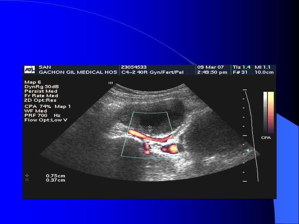
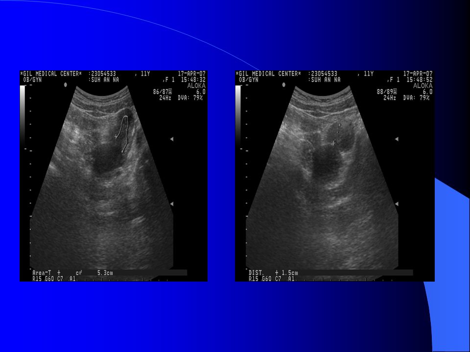
2007/3/9 * Uterus size: 7.3 X 4.5 X3.9 cm

12
RT.Ovary : 3.1 x 1.4 cm LT.Ovary : 4.3 x 5.6 cm

14
Uterus size: 8.8 X 2.8 cm / EM:0.58cm Two uterine horns. Obstruction of left side horn with hematometrocolpos.

15
IMP Uterus didelphys with left hematometrocolpos R/O Lt. ov cyst hydrosalpinx. R/O cystic teratoma 7.0x5.0cm sized low-level echoic region in Lt. ut

16
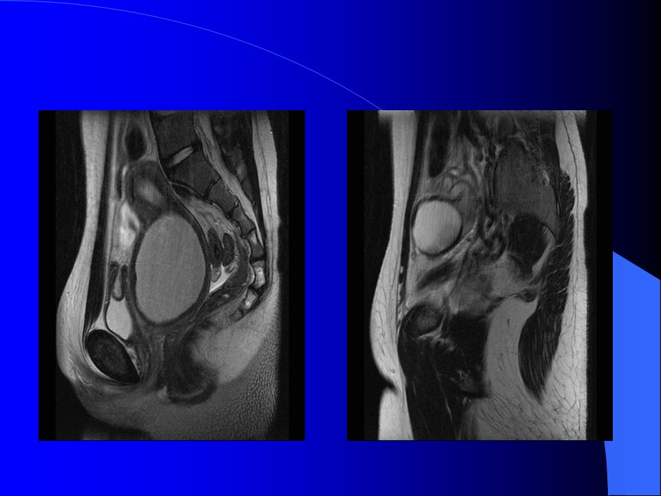
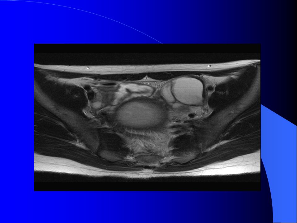
MRI 07/3/12

20
1. Absent left kidney with compensatory hypertrophy of the right kidney. 2. Two uterine horns (Uterine didelphys) Obstruction at left side, below cervical level with hematometrocolpos. -----> Mullerian duct dupilcation anomaly with unilateral kidney. 3. Dilated left salphinx with high signal fluid accumulation. ----> hydro- or hematosalphinx, left. 4. Normal appearing both ovaries.
 Obstruction at left side, below cervical level with hematometrocolpos > Mullerian duct dupilcation anomaly with unilateral kidney. 3. Dilated left salphinx with high signal fluid accumulation. ----> hydro- or hematosalphinx, left. 4. Normal appearing both ovaries..")
21
OP Findings OP date: 2007.3.15 Operation name : Lt. Cx. puncture & drainage Rt. Cx. : intact Lt. Cx. : 막혀 있어 puncture 후 old blood 약 100cc drainage 됨.

22
07/3/23 sono f/u * Uterus size: 7.3 X 4.5 X3.9 cm

23
RT.Ovary : 2.8 x 1.7 cm LT.Ovary : 3.6 x 2.0 cm

24
Uterus size: 7.9 X 4.5 X2.7cm EM:0.72cm

25
IMP: 1. Didelphys 2. Lt. ov. hemorrhagic mass.

26
07/04/17 sono f/u Uterus Size : rt:5.7 x 2.7 x 2.5 cm / lt: 9.8x2.8cm Endometrium : Rt:0.8 cm / Lt:0.6cm

29
RT.Ovary : 2.7 x 1.6 cm / LT.Ovary : 3.2 x 2.3 cm Lt. tube dilatation->thickness : 1.5cm. tube 안에 low-level echoic fluid collection 보입니다.

30
IMP: didelphys Lt. hydrosalpinx

31
Uterus didelphys with an obstructed hemivagina and ipsilateral renal agenesis 일측의 완전 혹은 불완전 질 폐쇄와 동측의 신장 무발생이 중복자궁과 동반된 증후군은 매우 드물다. ( Purslow, 1992) 여성에서 중복자궁이 있을 경우 43% 에서 일측 신장 무발 생이 동반되어 있고, 일측의 신장 무발생이 있을 경우 동반 되는 생식계통의 기형빈도는 50~70% 로 알려져 있다. (Perlmutter et al.,1986) 태생학적으로 이 증후군은 한쪽 중신관의 결손으로 인해 동측 신장과 요관이 발생하지 못하고 mullerian duct 의 부분적인 혹은 전체적인 융합의 이상에 의해 중복자궁이 형 성되어 발생한다. ( Marshall & Beisel, 1978;Maggee et al., 1979)
 여성에서 중복자궁이 있을 경우 43% 에서 일측 신장 무발 생이 동반되어 있고, 일측의 신장 무발생이 있을 경우 동반 되는 생식계통의 기형빈도는 50~70% 로 알려져 있다. (Perlmutter et al.,1986) 태생학적으로 이 증후군은 한쪽 중신관의 결손으로 인해 동측 신장과 요관이 발생하지 못하고 mullerian duct 의 부분적인 혹은 전체적인 융합의 이상에 의해 중복자궁이 형 성되어 발생한다. ( Marshall & Beisel, 1978;Maggee et al., 1979).")
32
태생 3 주경 어떤 기형발생원 이나 유전적 돌연변이에 의해 일측 pronephros 이 발생하지 않으면 이로 인해 동 측의 wolffian duct 혹은 metanephric duct 이 불완전 하 게 되어 동측의 신장이 형성되지 못한다. 그러나 태생 6 주경에 mullerian duct 는 형성되고 자라 나와 태생 8 주경에는 urogenital sinus 에서 만나게 된다. 하지만 urogenital sinus 에서 wolffian duct 의 불완전 형 성으로 mullerian ducts 의 융합은 일어나지 않게 되어 중복자궁이 생긴다. 질의 형성도 wolffian duct 의 영향을 받게 되는데 sinovaginal bulb 형성 시 wolffian duct 의 불완전 형성은 동측 질의 불완전 소통으로 폐쇄성 질을 유발한다고 알 려져 있다. (langman & wilson,1982)
.")
33
(nephrogenic ridge) ( uretral bud-wolffian duct)( metanephric blastema)
 ( uretral bud-wolffian duct)( metanephric blastema)")
34
The obstruction of one hemivagina will block outflow, resulting in complications such as hematocolpos, hematometra and hematosalpinx. The persistence of this situation may also be complicated by the occurrence of endometriosis as a result of blood reflux into the abdominal cavity. These anomalies are frequently accompanied by kidney and urinary tract malformations (i.e., kidney agenesis and dysplasia, double collecting system, ectopic ureter) on the same side as the defect. The manifesting symptoms usually appear only after menarche and consist of dysmenorrhea, severe abdominal pain, and the presence of an intraabdominal or pelvic mass.
 on the same side as the defect. The manifesting symptoms usually appear only after menarche and consist of dysmenorrhea, severe abdominal pain, and the presence of an intraabdominal or pelvic mass..")
Similar presentations


. CC: Low abd distension for 1 M CC: Low abd distension for 1 M (Pap: N these day, CA125: 15) (Pap: N these day, CA125: 15) Wt loss.>")


 김 응 록 정형외과 김 응 록 M.D, Ph, D>")


>")
 P(2) Att.: 우리 OBGY Fami. Hx : 시부 & 부 -> DM Px.Hx : (-) Why : Triple test(Edward syn) 초음파상 omphalocele,>")


>")