Download presentation
Presentation is loading. Please wait.

1
Gastric adenoma 에 대하여 ESD 시행 후 PDA 로 재발한 1 예 30th September 2009 Gastrointestinal Division of Internal Medicine Presbyterian Medical Center Young Jae Lee 대한소화기내시경학회 제 85 회 월례집담회

2
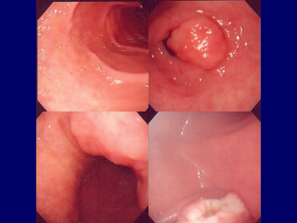
64/M Chief Complaint : G-ESD for known gastric adenoma

4
2005.5.12 Bx(#3) Tubular Adenoma with low grade dysplasia
 Tubular Adenoma with low grade dysplasia")
5
2005.5.30.ESD(1 st )
")
6
2005.5.31ESD(2 nd )
")
7
2005.6.2ESD(3 rd )
")
8
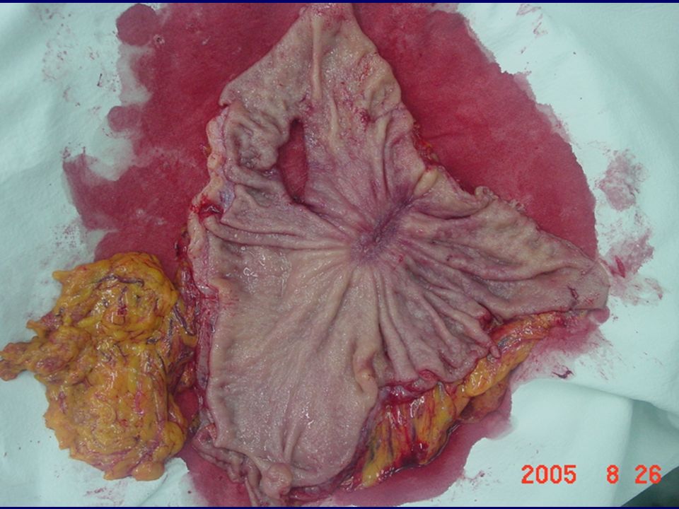
Pathologic Result 【 Gross 】 보내온 조직은 포르말린에 고정된 mucosal resection 된 위 점막 조직으로 크기는 7x5.5cm. 핀을 분리하였을 때 3 조각의 나뉘어져 있으며 큰 segment 가 가장 두꺼움. A1-3 : mucosa 가 가장 두꺼운 segment B1-3 : 가운데 segment C1-2 : 가장 작은 segment 【진단결과】 Stomach; endoscopic mucosal resection : 1.(A1-3) : Tubular adenoma with 1)severe atypia(High grade dysplasia) 2)involvement of lateral resection margin 3)clear basal resection margin 2.(B1-3)(C1-2) : Tubular adenoma with 1)moderate atypia(Low grade dysplasia) 2)involvement of lateral resection margin 3)clear basal resection margin
 : Tubular adenoma with 1)severe atypia(High grade dysplasia) 2)involvement of lateral resection margin 3)clear basal resection margin 2.(B1-3)(C1-2) : Tubular adenoma with 1)moderate atypia(Low grade dysplasia) 2)involvement of lateral resection margin 3)clear basal resection margin.")
10
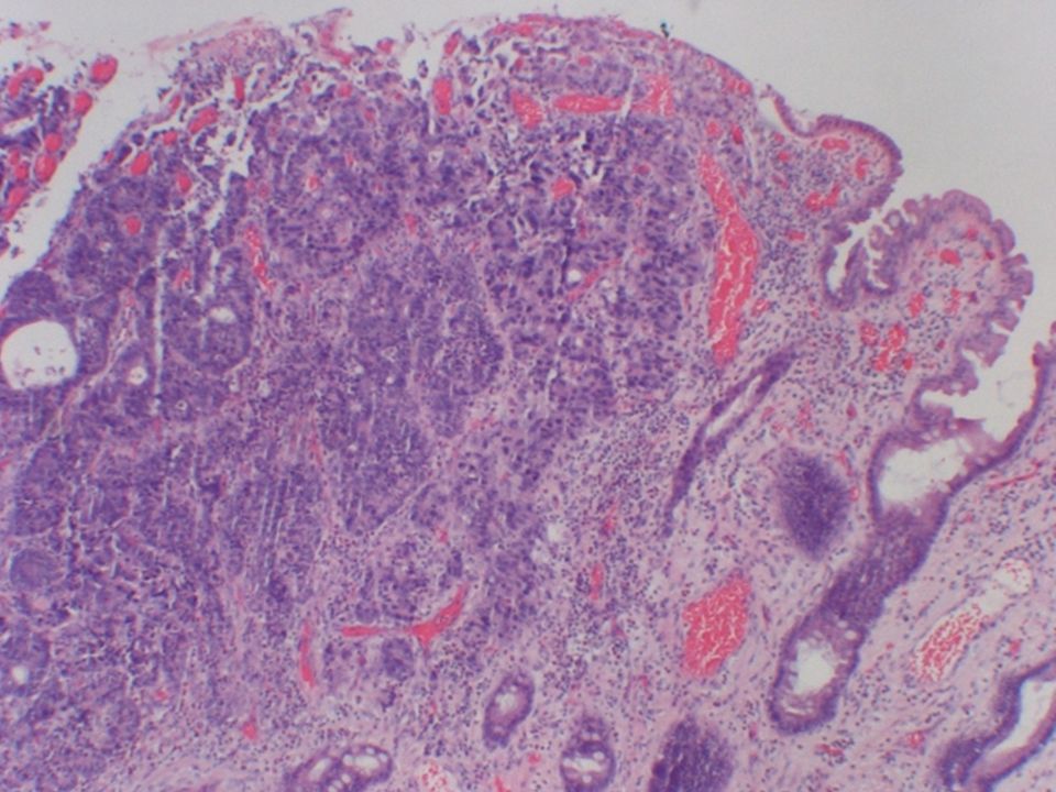
2005.8.10.f/u EGD ADENOCARCINOMA, poorly differentiated

14
Pathologic Result 【 Gross 】 보내온 조직은 포르말린에 고정되지 않은 부분 절제된 위장으로 크기는 소만을 따라 12cm, 대만을 따라 16cm. 체부의 전벽에 gastrotomy 흔적이 있으며 길이는 4cm. 대만곡을 따라 열었을 때 체부의 소만곡에 EMR 흔적이 있으며 분명한 종괴는 보이지 않음. EMR 흔적은 upper resection margin 에서 1cm, lower resection margin 에서 8cm 떨어져 있음. F/S:upper resection margin A1-7:EMR site B:lower resection margin C1-10:lymph nodes 【진단결과】 Stomach, radical subtotal gastrectomy : TUBULAR ADENOCARCINOMA, poorly differentiated with 1) tumor size : 직경 1cm. 2) confined to mucosa. 3) no definitive lymphovascular invasion. 4) clear resection margins. 5) lymphocytic infiltration, grade 2/3. 6) greater curvature lymph node:free of carcinoma(0/2). lesser curvature lymph node:free of carcinoma(0/4). common hepatic lymph node:free of carcinoma(0/5). celiac lymph node:free of carcinoma(0/2). splenic lymph node:free of carcinoma(0/1). left gastric lymph node:free of carcinoma(0/2). right paracardiac lymph node:free of carcinoma(0/9). suprapyloric lymph node:free of carcinoma(0/1). infrapyloric lymph node:free of carcinoma(0/4). hepatoduodenal lymph node:free of carcinoma(0/2).
 tumor size : 직경 1cm. 2) confined to mucosa. 3) no definitive lymphovascular invasion. 4) clear resection margins. 5) lymphocytic infiltration, grade 2/3. 6) greater curvature lymph node:free of carcinoma(0/2). lesser curvature lymph node:free of carcinoma(0/4). common hepatic lymph node:free of carcinoma(0/5). celiac lymph node:free of carcinoma(0/2). splenic lymph node:free of carcinoma(0/1). left gastric lymph node:free of carcinoma(0/2). right paracardiac lymph node:free of carcinoma(0/9). suprapyloric lymph node:free of carcinoma(0/1). infrapyloric lymph node:free of carcinoma(0/4). hepatoduodenal lymph node:free of carcinoma(0/2)..")
15
Discrepancy of the histopathological diagnosis Endoscopic forcep biopsy Endscopic submucosal dissection 대한소화기내시경학회지 2009:38:188-192

16
Is ESD pathologic result accurate? Risk factors associated with local recurrence 1)Incomplete resection 2)Tumor size & location 3)Piecemeal resection Gastrointest Endosc. 2008 Nov;68(5):887-94 대한소화기내시경학회지 2007:35:6-13 Specimen related factor? 1)Electrocauterization 2)Autolysis 대한소화기내시경학회지 2008;37(Suppl. 2):134 Unknown factor? ESD : adenoma with HG →? → PDA recurrence
Incomplete resection 2)Tumor size & location 3)Piecemeal resection Gastrointest Endosc Nov;68(5): 대한소화기내시경학회지 2007:35:6-13 Specimen related factor. 1)Electrocauterization 2)Autolysis 대한소화기내시경학회지 2008;37(Suppl. 2):134 Unknown factor. ESD : adenoma with HG →. → PDA recurrence.")
Similar presentations


 유명한 갯벌 ( 우리나라 ), 여러 갯벌 축제 갯벌이 만들어지는 조건 람사르 협약이란 ? 람사르 협약에 가입된 우리나라 생태지 밀물과 썰물 갯벌에.>")
 현장조사, 의식확인, 연락 현장은 안전한가 조사한다. 119 나 응급의료기관에 연락한다. 발바닥을 간지럽히거나 가볍게 꼬집어 본다. 0 ~ 4 분 4 ~ 6 분 6 ~ 10 분 10.>")


 발표자 : 농어업조사과 장 천 숙. 목 차 1 월별 작업 흐름 2 재배 방법 3 병충해 방지 4 수박의 효능.>")


 )>")


