Download presentation
Presentation is loading. Please wait.

2
Peripheral Nervous System Chapter 6 Copyright The McGraw-Hill Companies, Inc. Permission required for reproduction or display.

6
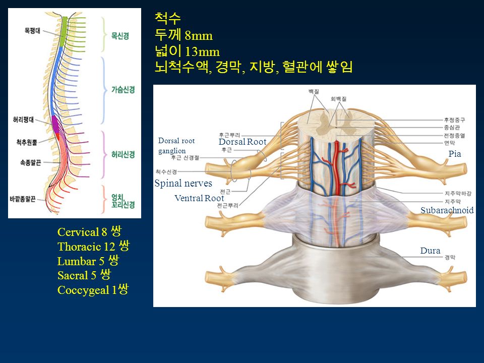
척수 두께 8mm 넓이 13mm 뇌척수액, 경막, 지방, 혈관에 쌓임 Cervical 8 쌍 Thoracic 12 쌍 Lumbar 5 쌍 Sacral 5 쌍 Coccygeal 1 쌍 Spinal nerves Dorsal root ganglion Dorsal Root Ventral Root Dura Pia Subarachnoid

7
Plexus 신경총 Cervical 경신경총 Brachial 완신경총 Lumbar enlargement ( 요팽대 ) Sacral 천신경총 神經叢
 Sacral 천신경총 神經叢")
10
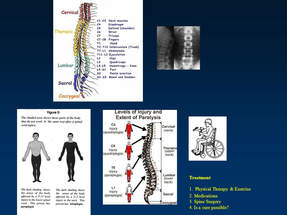
Treatment 1. Physical Therapy & Exercise 2. Medications 3. Spine Surgery 4. Is a cure possible?

11
Peripheral Nervous System 말초신경계末梢神經系 감각 CNS 운동 : 항상성 유지 중추와 신체간의 빠른 정보소통 구성 : –Cranial nerves 뇌신경腦神經 12 쌍 –Spinal nerve 척수신경脊髓神經 31 쌍 대부분 감각과 운동신경세포의 축삭 ( 뇌신경 3 은 예외 )
")
13
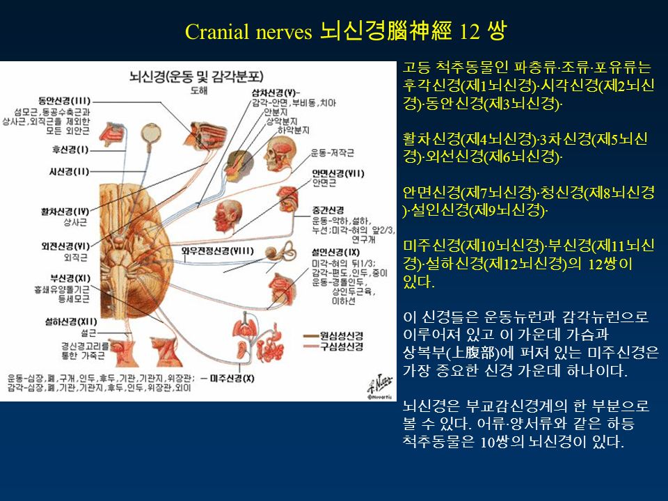
고등 척추동물인 파충류 · 조류 · 포유류는 후각신경 ( 제 1 뇌신경 )· 시각신경 ( 제 2 뇌신 경 )· 동안신경 ( 제 3 뇌신경 )· 활차신경 ( 제 4 뇌신경 )·3 차신경 ( 제 5 뇌신 경 )· 외선신경 ( 제 6 뇌신경 )· 안면신경 ( 제 7 뇌신경 )· 청신경 ( 제 8 뇌신경 )· 설인신경 ( 제 9 뇌신경 )· 미주신경 ( 제 10 뇌신경 )· 부신경 ( 제 11 뇌신 경 )· 설하신경 ( 제 12 뇌신경 ) 의 12 쌍이 있다. 이 신경들은 운동뉴런과 감각뉴런으로 이루어져 있고 이 가운데 가슴과 상복부 ( 上腹部 ) 에 퍼져 있는 미주신경은 가장 중요한 신경 가운데 하나이다. 뇌신경은 부교감신경계의 한 부분으로 볼 수 있다. 어류 · 양서류와 같은 하등 척추동물은 10 쌍의 뇌신경이 있다. Cranial nerves 뇌신경腦神經 12 쌍
 에 퍼져 있는 미주신경은 가장 중요한 신경 가운데 하나이다. 뇌신경은 부교감신경계의 한 부분으로 볼 수 있다. 어류 · 양서류와 같은 하등 척추동물은 10 쌍의 뇌신경이 있다. Cranial nerves 뇌신경腦神經 12 쌍.")
14
눈돌림신경 도르래신경 갓돌림신경 얼굴신경 속귀신경 혀밑신경 혀의 운동목과 등 일부분 운동 가장 길고 광범위 ( 소화, 순환, 호흡 등 내장운동 ) 청각, 평형감각 얼굴, 목의 근육조절 혀 앞쪽 2/3 맛
 청각, 평형감각 얼굴, 목의 근육조절 혀 앞쪽 2/3 맛")
15
Spinal nerve 척수신경脊髓神經 31 쌍

16
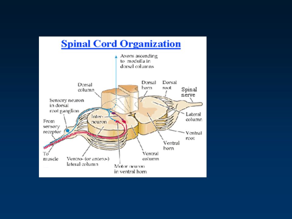
Spinal Nerve – Spinal Cord Interface Spinal nerves join with spinal cord through spinal nerve roots –Dorsal Root Carry afferent axons into the spinal cord Dorsal root ganglion –Contains afferent cell bodies –Ventral Root Carry efferent axons out of spinal cord Sensory neurons synapse with motor and association neurons Motor neurons stimulated by association neurons or directly by sensory neurons Association neurons allow both reflexive activity and motor control by upper motor neurons –Assoc. neurons of descending tracts that originate in the brain Somatic motor neurons = lower motor neurons Fig 6.2

19
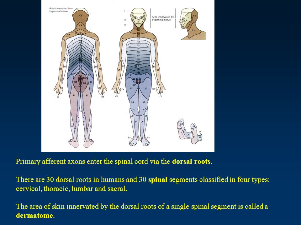
Primary afferent axons enter the spinal cord via the dorsal roots. There are 30 dorsal roots in humans and 30 spinal segments classified in four types: cervical, thoracic, lumbar and sacral. The area of skin innervated by the dorsal roots of a single spinal segment is called a dermatome.

23
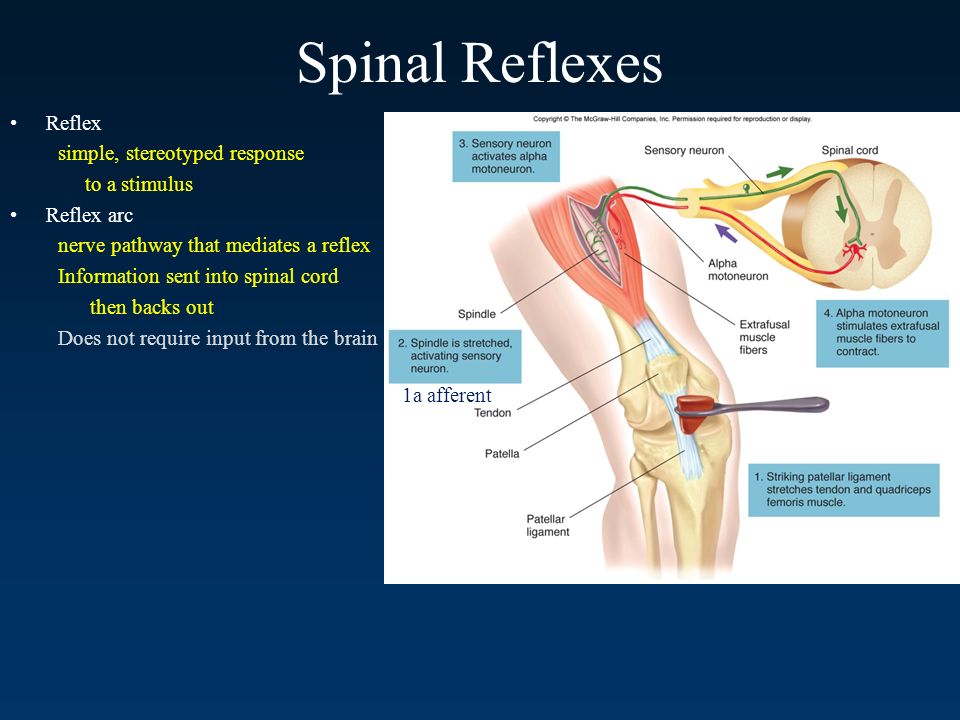
Spinal Reflexes Reflex simple, stereotyped response to a stimulus Reflex arc nerve pathway that mediates a reflex Information sent into spinal cord then backs out Does not require input from the brain Fig 6.3 1a afferent

24
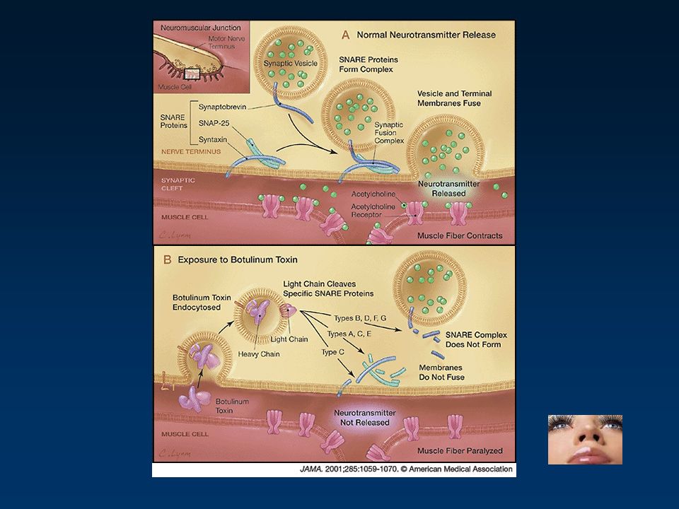
The motor unit = a motor neuron + the muscle fibers it innervates. A muscle cell is innervated by only one neuron. An alpha motor neuron may innervate many muscle fibers (3- 2000). The fewer fibers involved, the finer the muscle control will be. Cell bodies for the alpha motor neurons are located in the spinal cord. Acetylcholine is the neurotransmitter between the motor neuron and the muscle cell, and the muscle cell has nicotinic receptors.
. The fewer fibers involved, the finer the muscle control will be. Cell bodies for the alpha motor neurons are located in the spinal cord. Acetylcholine is the neurotransmitter between the motor neuron and the muscle cell, and the muscle cell has nicotinic receptors..")
31
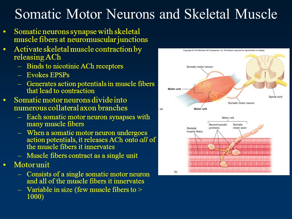
Somatic Motor Neurons and Skeletal Muscle Somatic neurons synapse with skeletal muscle fibers at neuromuscular junctions Activate skeletal muscle contraction by releasing ACh –Binds to nicotinic ACh receptors –Evokes EPSPs –Generates action potentials in muscle fibers that lead to contraction Somatic motor neurons divide into numerous collateral axon branches –Each somatic motor neuron synapses with many muscle fibers –When a somatic motor neuron undergoes action potentials, it releases ACh onto all of the muscle fibers it innervates –Muscle fibers contract as a single unit Motor unit –Consists of a single somatic motor neuron and all of the muscle fibers it innervates –Variable in size (few muscle fibers to > 1000) Fig 6.6
 Fig 6.6")
33
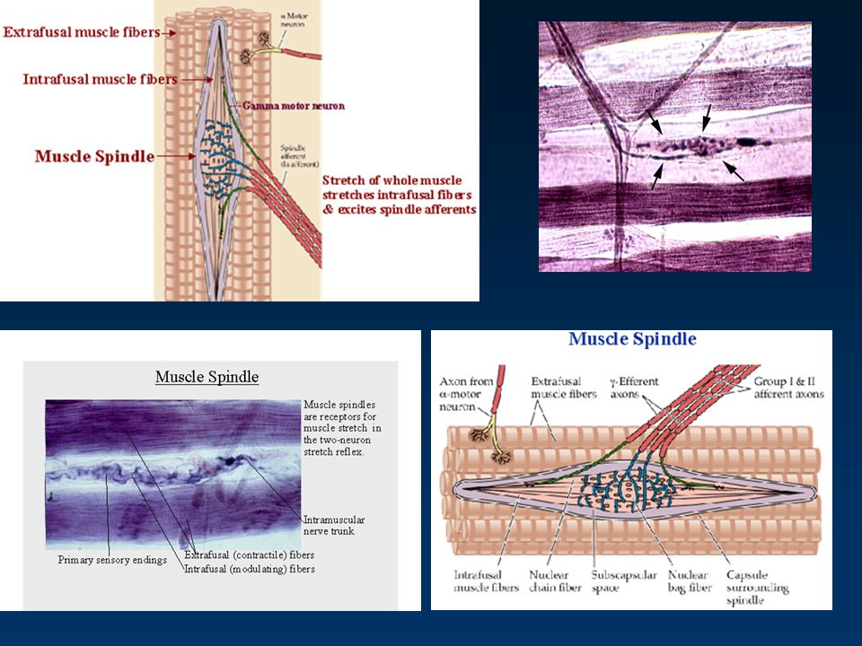
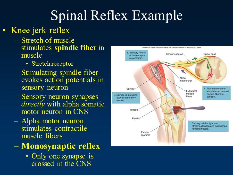
Spinal Reflex Example Knee-jerk reflex –Stretch of muscle stimulates spindle fiber in muscle Stretch receptor –Stimulating spindle fiber evokes action potentials in sensory neuron –Sensory neuron synapses directly with alpha somatic motor neuron in CNS –Alpha motor neuron stimulates contractile muscle fibers –Monosynaptic reflex Only one synapse is crossed in the CNS Fig 6.3

36
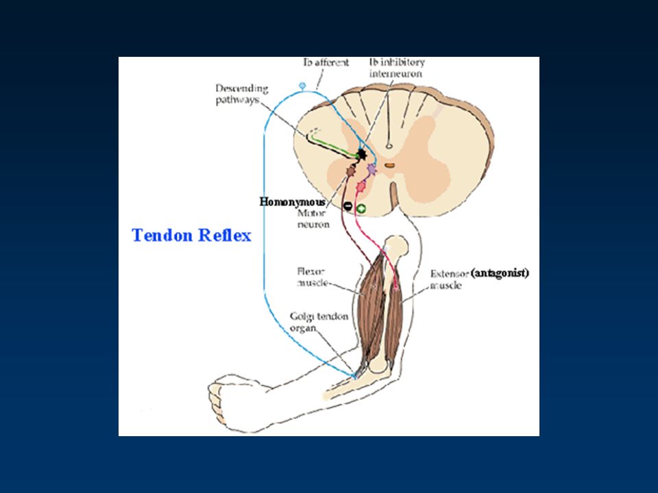
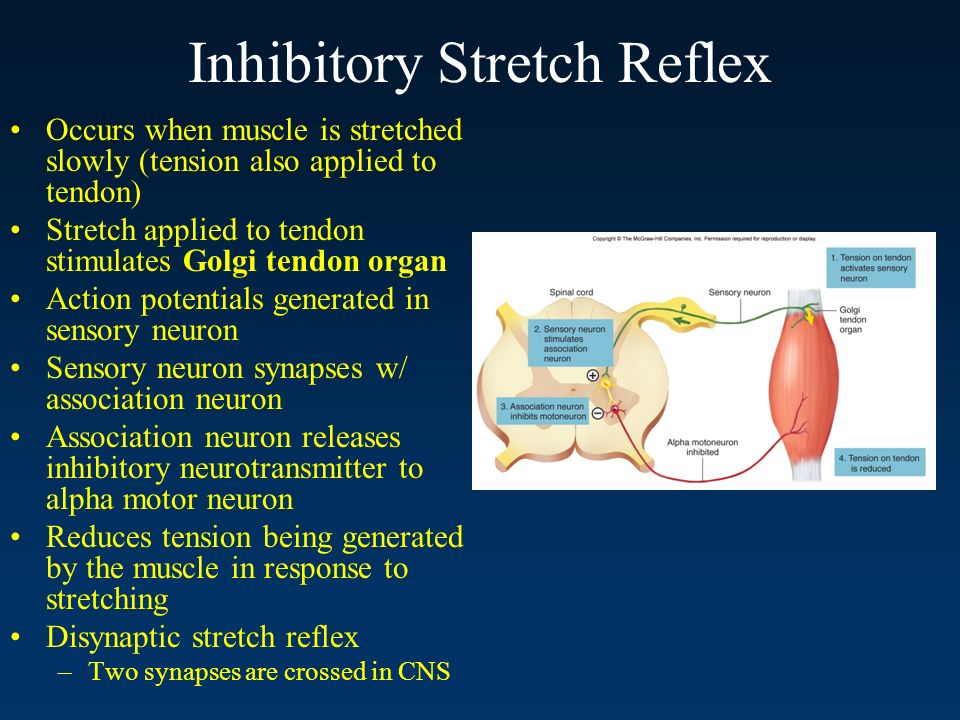
Inhibitory Stretch Reflex Occurs when muscle is stretched slowly (tension also applied to tendon) Stretch applied to tendon stimulates Golgi tendon organ Action potentials generated in sensory neuron Sensory neuron synapses w/ association neuron Association neuron releases inhibitory neurotransmitter to alpha motor neuron Reduces tension being generated by the muscle in response to stretching Disynaptic stretch reflex –Two synapses are crossed in CNS Fig 6.4
 Stretch applied to tendon stimulates Golgi tendon organ Action potentials generated in sensory neuron Sensory neuron synapses w/ association neuron Association neuron releases inhibitory neurotransmitter to alpha motor neuron Reduces tension being generated by the muscle in response to stretching Disynaptic stretch reflex –Two synapses are crossed in CNS Fig 6.4")
39
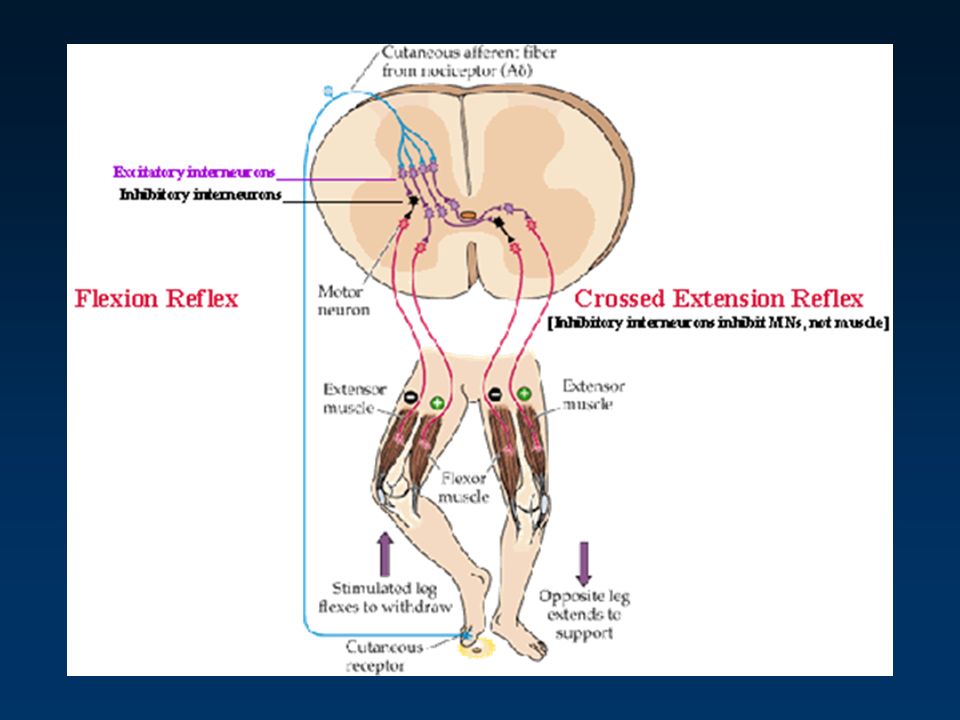
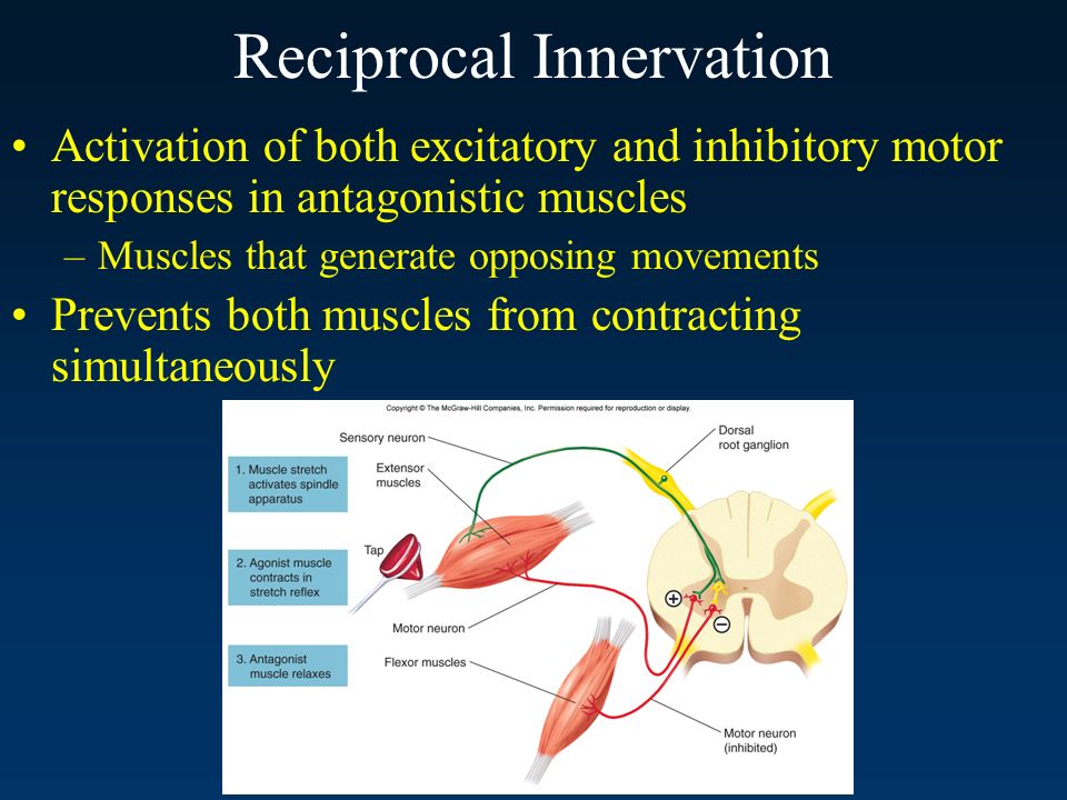
Reciprocal Innervation Activation of both excitatory and inhibitory motor responses in antagonistic muscles –Muscles that generate opposing movements Prevents both muscles from contracting simultaneously

41
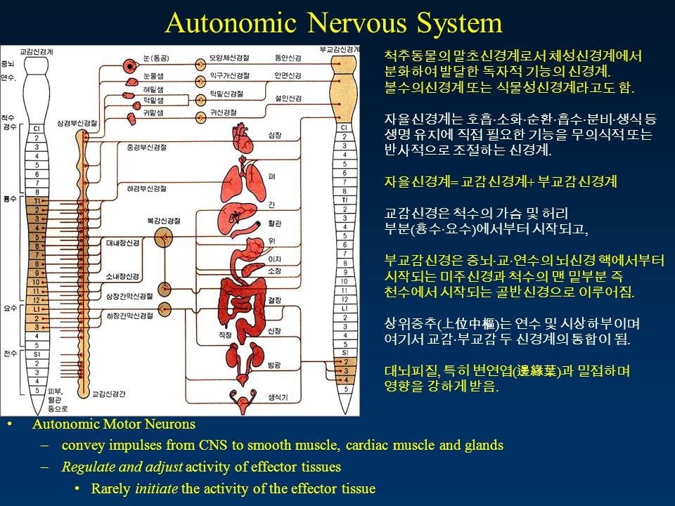
척추동물의 말초신경계로서 체성신경계에서 분화하여 발달한 독자적 기능의 신경계. 불수의신경계 또는 식물성신경계라고도 함. 자율신경계는 호흡 · 소화 · 순환 · 흡수 · 분비 · 생식 등 생명 유지에 직접 필요한 기능을 무의식적 또는 반사적으로 조절하는 신경계. 자율신경계 = 교감신경계 + 부교감신경계 교감신경은 척수의 가슴 및 허리 부분 ( 흉수 · 요수 ) 에서부터 시작되고, 부교감신경은 중뇌 · 교 · 연수의 뇌신경 핵에서부터 시작되는 미주신경과 척수의 맨 밑부분 즉 천수에서 시작되는 골반신경으로 이루어짐. 상위중추 ( 上位中樞 ) 는 연수 및 시상하부이며 여기서 교감 · 부교감 두 신경계의 통합이 됨. 대뇌피질, 특히 변연엽 ( 邊緣葉 ) 과 밀접하며 영향을 강하게 받음. Autonomic Nervous System Autonomic Motor Neurons –convey impulses from CNS to smooth muscle, cardiac muscle and glands –Regulate and adjust activity of effector tissues Rarely initiate the activity of the effector tissue
 에서부터 시작되고, 부교감신경은 중뇌 · 교 · 연수의 뇌신경 핵에서부터 시작되는 미주신경과 척수의 맨 밑부분 즉 천수에서 시작되는 골반신경으로 이루어짐. 상위중추 ( 上位中樞 ) 는 연수 및 시상하부이며 여기서 교감 · 부교감 두 신경계의 통합이 됨. 대뇌피질, 특히 변연엽 ( 邊緣葉 ) 과 밀접하며 영향을 강하게 받음. Autonomic Nervous System Autonomic Motor Neurons –convey impulses from CNS to smooth muscle, cardiac muscle and glands –Regulate and adjust activity of effector tissues Rarely initiate the activity of the effector tissue.")
42
Autonomic Efferent Pathways Two neurons involved in efferent pathway (connecting CNS to effector) 1st (preganglionic) has cell body in CNS –synapses with 2nd in the autonomic ganglion 2nd (postganglionic) sends signal from auton. gang. to the effector organ NOTE – somatic efferent pathways have only a single motor neuron extending from the CNS to the skeletal muscle fibers Fig 6.7

43
교감신경은 12 쌍의 가슴신경과 1, 2 번째 허리신경의 일부에 의해 구성됨. 척추옆에서 교감신경절을 만들고 여기서부터 신체 각부위로 퍼져나감. 부교감신경은 3, 7, 9, 10 번째 뇌신경과 2, 3, 4 번째 엉치신경의 일부에 의해 이루어짐. 지배하는 장기 근처에서 부교감신경절을 만들고 여기에서 각 장기로 퍼져 나감. 그 중 미주신경이 가장 넓은 부위를 담당하지.

44
Autonomic Nervous System: Sympathetic Division Sympathetic (thoracolumnar) –Preganglionic axons enter sympathetic chain of ganglia just lateral to spinal cord –Ganglia in chain linked via collateral axons branching from preganglionic motor neurons –Usually synapse with postganglionic motor neurons in the chain
 –Preganglionic axons enter sympathetic chain of ganglia just lateral to spinal cord –Ganglia in chain linked via collateral axons branching from preganglionic motor neurons –Usually synapse with postganglionic motor neurons in the chain")
45
교감신경계 (sympathetic division) 의 다른 이름은 thoracolumbar division 세포체는 척수의 흉부 분절 (thoracic segment) 과 요부 분절 (lumbar segment) 에 위치. 섬유들의 축삭 (axon) 은 척수관의 바로 바깥에 위치하는 두 chain 에 대부분 위치하는 교감신경절 (sympathetic ganglia). 절 안에서 절전섬유와 절후섬유는 시냅스 ---> 절후 축삭은 내장 효과기에 하나의 절전섬유는 종종 많은 효과기로 가는 여러 절후섬유와 시냅스를 이룸. 교감신경계는 많은 장기에서 광범위한 반응. 교감신경계는 운동이나 스트레스 상황 - 분노, 공포, 불안 등등 - 에서 지배적. 심박수는 증가하고 골격근의 혈관확장은 많은 산소를 공급 세기관지 (bronchiole) 의 확장은 많은 산소를 얻게 간은 글리코겐을 에너지원으로 쓰이는 포도당으로 변환 소화액 분비는 감소하고 장연동운동은 느려짐. 피부와 내부장기의 혈관수축은 중요기관 ( 뇌, 간, 심장 등 ) 으로 더 많은 피를 보낼 수 있게 Sympathetic Division
 은 척수관의 바로 바깥에 위치하는 두 chain 에 대부분 위치하는 교감신경절 (sympathetic ganglia). 절 안에서 절전섬유와 절후섬유는 시냅스 ---> 절후 축삭은 내장 효과기에 하나의 절전섬유는 종종 많은 효과기로 가는 여러 절후섬유와 시냅스를 이룸. 교감신경계는 많은 장기에서 광범위한 반응. 교감신경계는 운동이나 스트레스 상황 - 분노, 공포, 불안 등등 - 에서 지배적. 심박수는 증가하고 골격근의 혈관확장은 많은 산소를 공급 세기관지 (bronchiole) 의 확장은 많은 산소를 얻게 간은 글리코겐을 에너지원으로 쓰이는 포도당으로 변환 소화액 분비는 감소하고 장연동운동은 느려짐. 피부와 내부장기의 혈관수축은 중요기관 ( 뇌, 간, 심장 등 ) 으로 더 많은 피를 보낼 수 있게 Sympathetic Division.")
46
Autonomic Nervous System: Parasympathetic Division Parasympathetic (craniosacral) –Preganglionic axons synapse with postganglionic motor neurons at terminal ganglia (adjacent to or within target organ) Fig 6.10
 –Preganglionic axons synapse with postganglionic motor neurons at terminal ganglia (adjacent to or within target organ) Fig 6.10")
47
부교감신경계 (parasympathetic division) 의 다른 이름은 craniosacral division. 부교감신경 절전 섬유의 세포체는 뇌간 (brain stem) 과 척수의 미부분절 (sacral segment) 에 위치. 축삭은 3,7,9,10 번 뇌신경과 몇몇 미부 신경에 있으며 부교감 신경절까지 다다름. 절들은 내장 효과기에 근접하거나 그 안에 위치. 효과기의 세포까지 매우 짧은 축삭을 가진 세포체. 하나의 절전 섬유는 하나의 효과기로만 가는 소수의 절후섬유와 시냅스. 이러한 해부학적 배열로 인해 매우 국소적인 ( 한 장기에 국한된 ) 반응. 부교감신경계는 이완 ( 비스트레스 ) 된 상황에서 정상적 장기기능을 항진. 소화는 소화액분비가 증진되고 연동운동이 항진되어 효과적으로 일어남. 배변과 배뇨가 일어남. 심박수는 정상 휴식기의 수치로 유지. Parasympathetic Division
 과 척수의 미부분절 (sacral segment) 에 위치. 축삭은 3,7,9,10 번 뇌신경과 몇몇 미부 신경에 있으며 부교감 신경절까지 다다름. 절들은 내장 효과기에 근접하거나 그 안에 위치. 효과기의 세포까지 매우 짧은 축삭을 가진 세포체. 하나의 절전 섬유는 하나의 효과기로만 가는 소수의 절후섬유와 시냅스. 이러한 해부학적 배열로 인해 매우 국소적인 ( 한 장기에 국한된 ) 반응. 부교감신경계는 이완 ( 비스트레스 ) 된 상황에서 정상적 장기기능을 항진. 소화는 소화액분비가 증진되고 연동운동이 항진되어 효과적으로 일어남. 배변과 배뇨가 일어남. 심박수는 정상 휴식기의 수치로 유지. Parasympathetic Division.")
48
Sympathetic vs. Parasympathetic Effects Sympathetic –dominates in stressful situations –prepares body for activity (“Fight or Flight”) –adrenergic effects Most postganglionic sympathetic neurons release norepinephrine onto the effector tissues Effector tissues may have different forms of adrenergic receptors ( α or β, with different subvariants)
 –adrenergic effects Most postganglionic sympathetic neurons release norepinephrine onto the effector tissues Effector tissues may have different forms of adrenergic receptors ( α or β, with different subvariants).")
49
교감신경이 자극됐을 때의 효과는 사람이 아주 긴장했을 때 나타나는 반응 싸움을 할 때의 신체반응 심장은 빨리 뛰어 근육으로 많은 피를 내보내고 호흡기는 넓어져서 숨을 많이 쉴 수 있고 눈동자가 커져서 상대방을 잘 볼 수 있게 되며 싸움에 당장 필요 없는 소화관의 운동은 저하됨.

50
Sympathetic vs. Parasympathetic Effects Parasympathetic –dominates during relaxed situations –precise control over the body (“Rest and Digest”) –cholinergic effects Most postganglionic sympathetic neurons release acetylcholine onto the effector tissues Effector tissues possess muscarinic ACh receptors Table 6.2
 –cholinergic effects Most postganglionic sympathetic neurons release acetylcholine onto the effector tissues Effector tissues possess muscarinic ACh receptors Table 6.2.")
Similar presentations

. Why must organisms reproduce?>")


 인체기능 – 생리학 (Physiology) 생리학 인체의 세포, 조직 또는 각 기관이나 계통 등과 같은 인체구성단위 들이 생명현상을.>")


 해부생리학의 정의 해부생리학 해부학(anatomy) :인체 기관의 구조와 이들 구조가 어떻게 상호 관련하는 지를 연구 하는 학문 생리학(physiology)>")
>")


>")
