Download presentation
Presentation is loading. Please wait.

1
콩팥종양, 고환 및 기타 종양 상부요로종양, 방광종양
순천향대학교 비뇨기학교실 교수 양원재 2016년 1학기 신장요로생식학

2
2013년 국가암정보센터

4
강의계획 1. Renal cell carcinoma 2. Urothelial cancer Bladder Renal pelvis
Ureter 3. Testis, Penis Cancer

5
Cystic renal masses A renal mass that is not clearly a simple cyst by strict ultrasound criteria should be evaluated further with computed tomography (CT). A dedicated (thin-slice) renal CT scan remains the single most important radiographic test for delineating the nature of a renal mass.
. A dedicated (thin-slice) renal CT scan remains the single most important radiographic test for delineating the nature of a renal mass.")
6
Solid

7
Needle aspiration 시 cyst가 터지면 cancer cell seeding Bx 불가..

8
Bosniak II

9
Bosniak III

10
Bosnik IV

11
Solid renal masses

12
Clinical Presentation
Because of the sequestered location of the kidney within the retroperitoneum, many renal masses remain asymptomatic and nonpalpable until they are advanced. With the more pervasive use of noninvasive imaging for the evaluation of a variety of nonspecific symptom complexes, more than 50% of RCCs are now detected incidentally.

13
Symptomatic renal mass
동영상

14
Symptomatic renal mass
동영상
15
Tx for localized RCC Radical or partial nephrectomy

16
Postoperative nomogram predicting recurrence of clear cell renal cell
72점 40점 28점 Postoperative nomogram predicting recurrence of clear cell renal cell carcinoma after nephrectomy

17
Tx for mRCC

18
Surgical Management of mRCC -Debulking or Cytoreductive Nephrectomy-
Targeted molecular agents in Clear Cell mRCC -Antagonists of the Vascular Endothelial Growth Factor Pathway-

19
Motzer RJ, et al. N Engl J Med 2007;356:115–24

20
요약 초음파에서 단순 낭종 이외의 신종물이 관찰될 때에는 조영증 강 CT를 시행해야 한다.
낭성 신종물에 대한 Bosniak 분류에서 그룹 III 이상은 외과적 적출이 필요한 대상이다. 국소 신세포암은 외과적 절제가 최선이며 크기가 작을 경우 콩 팥 전체를 제거하지 않고 암종만 제거해도 된다. 전이 신세포암에서 가능한 원발암을 제거하는 것이 추후 전신 치료에 대한 반응을 높혀준다. 전이 신세포암은 기존의 고식적인 항암, 방사선 치료에 반응하 지 않고 표적치료가 표준적인 치료이다.

21
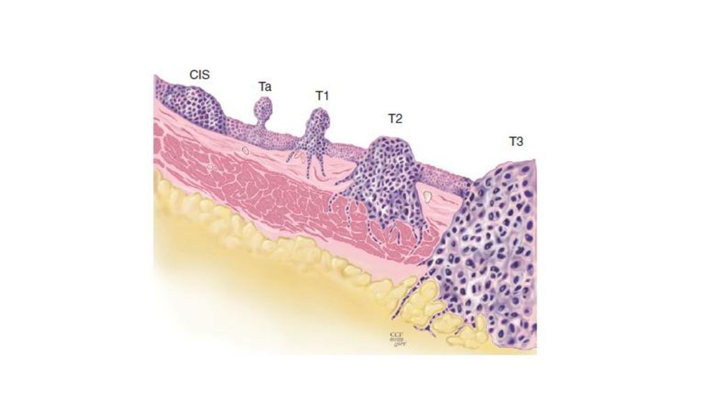
Urothelial Cancer: Bladder
Endoscopic surgery, BCG instillation Radical surgery

23
Detection of Urothelial Carcinoma
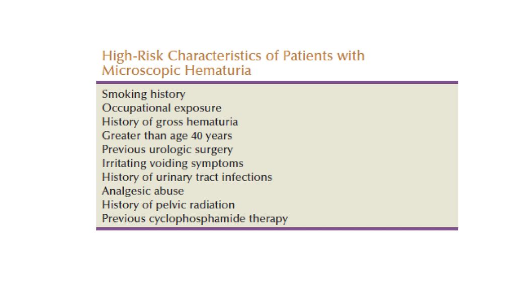
Gross, painless hematuria is the primary symptom in 85% of patients with a newly diagnosed bladder tumor, and microscopic hematuria occurs in virtually all patients. The hematuria is usually intermittent; therefore any episode of gross hematuria should be evaluated even if subsequent urinalysis is negative. Fifty percent of patients with gross hematuria will have a demonstrable cause, 20% will have a urologic malignancy, and 12% will have a bladder tumor.

25
A full hematuria evaluation
Cystoscopy Urine cytology Upper-tract imaging (primarily a CT scan of the abdomen and pelvis) PSA blood test
 PSA blood test.")
26
TransUrethral Resection of Bladder tumor –TURBt -
(1) remove all visible tumors (2) provide specimens for pathologic examination to determine stage and grade.
 remove all visible tumors (2) provide specimens for pathologic examination to determine stage and grade.")
27
Single papillary mass 동영상

28
Multiple papillary mass
동영상

29
Multiple solid mass 동영상

30
Advanced Bladder Urothelial cancer
동영상

31
Radical cystoprostatectomy
Included; Bladder Perivesical fat Peritoneum Lower ureters Prostate Seminal vesicle
32
Bilateral Pelvic Lymph Nodes Dissection - Therapeutic procedure -
33
Ileal conduit, Urostomy, 요루
34
Orthotopic Neobladder

35
Orthotopic ileal neobladder substitution: Studer neobladder

36
Urothelial Cancer: Renal pelvis

37
Incidentally found Rt hydronephrosis, GIII

38
Retrograde pyelography: Renal pelvis mass
동영상

39
CT: Renal pelvis tumor 동영상

40
Urothelial Cancer: Ureter
동영상

41
Testis cancer Germ cell tumor Lymphoid and Hematopoietic Tumors
Lymphoma Plasmacytoma Leukemia

42
Testis Cancer: Germ Cell Tumor

43
Initial presentation Painless testicular mass
A firm intratesticular mass should be considered cancer until proven otherwise and should be evaluated further with scrotal ultrasonography. Prior studies show that up to one third of testicular tumors are initially misdiagnosed as epididymitis or hydrocele.

44
Diagnosis In men presenting with a testicular mass, hydrocele, or unexplained scrotal symptoms or signs, scrotal ultrasonography should be considered an extension of the physical examination because it is widely available, inexpensive, and noninvasive. Testicular cancer is one of the few malignancies associated with accurate serum tumor markers (LDH, AFP, and hCG), a finding that is essential in its diagnosis, prognosis, treatment, and monitoring. Serum tumor marker levels should be obtained at diagnosis, after orchiectomy, to monitor for response to chemotherapy, and to monitor for relapse in patients on surveillance and after completion of therapy.
, a finding that is essential in its diagnosis, prognosis, treatment, and monitoring. Serum tumor marker levels should be obtained at diagnosis, after orchiectomy, to monitor for response to chemotherapy, and to monitor for relapse in patients on surveillance and after completion of therapy.")
45
Scrotal sonography 동영상

46
Tx: Seminomatous GCT

47
Tx: Non-Seminomatous GCT

48
RetroPeritoneal Lymph Node Dissection: RPLND

49
John P. Donohue Lawrence Einhorn Their works led to an increase in cure rate of testicular cancer from 5% to 90%.

50
Penile Cancer
Similar presentations
. CC: Low abd distension for 1 M CC: Low abd distension for 1 M (Pap: N these day, CA125: 15) (Pap: N these day, CA125: 15) Wt loss.>")


, PR(+) –HER-2: +3.>")


 주요 내용 소개>")
 1.>")


>")


>")


