Download presentation
Presentation is loading. Please wait.

1
Nervous System Neurons and Synapses
Chapter 4 Copyright The McGraw-Hill Companies, Inc. Permission required for reproduction or display.

2
Nervous System神經系: General Organization
申亨澈 敎授
3
Nervous System神經系: General Organization
Central Nervous System (CNS 중추신경계) Brain (腦)+ Spinal Cord(脊髓) Control + integration Peripheral Nervous System (PNS 말초신경계) cranial nerves(뇌신경) and spinal nerves(척수신경) Communication connects CNS to sensory receptors, muscles and glands 구심성 (afferent)감각신경세포 수의적 원심성 (efferent)운동신경세포 불수의적 자율운동신경세포 교감, 부교감자율신경세포
 Brain (腦)+ Spinal Cord(脊髓) Control + integration. Peripheral Nervous System (PNS 말초신경계) cranial nerves(뇌신경) and spinal nerves(척수신경) Communication. connects CNS to sensory receptors, muscles and glands. 구심성 (afferent)감각신경세포. 수의적. 원심성 (efferent)운동신경세포. 불수의적. 자율운동신경세포. 교감, 부교감자율신경세포.")
4
Cell Types세포종류 Neurons 신경細胞 Neuroglia (glia) 신경교 神經膠 교膠
目, 耳 感覺신경 運動, 연합신경세포 의사단극擬似單極 쌍극雙極 다극多極 축색軸索 수상돌기 樹狀突起 Neurons 신경細胞 conduct electrical signals 전기신호의 전도 電氣信號의 傳導 Neuroglia (glia) 신경교 神經膠 교膠 Majority of all nerve tissue cells support neurons 성상星狀교세포 희소돌기 아교세포 수초 髓鞘 뇌실막세포 뇌척수액 미세아교세포
 신경교 神經膠 교膠. Majority of all nerve tissue cells. support neurons. 성상星狀교세포. 희소돌기 아교세포. 수초 髓鞘. 뇌실막세포. 뇌척수액. 미세아교세포.")
5
Neuron Structure Cell Body 細胞體 세포체 Dendrites樹狀突起 수상돌기 Axon
Nucleus (核) and typical organelles (세포小器官) Dendrites樹狀突起 수상돌기 Receive stimulation Axon Conduct electrical signals (action potentials 활동전위) Often insulated with a myelin sheath Axon hillock site where AP’s originate Axon terminals 축삭終末 where chemical signals are released 축색구 軸索 축색 軸索 축색 수상돌기 축색구 無髓영역 有髓영역 란비에 결절 수초 髓鞘
 and typical organelles (세포小器官) Dendrites樹狀突起 수상돌기. Receive stimulation. Axon. Conduct electrical signals (action potentials 활동전위) Often insulated with a myelin sheath. Axon hillock. site where AP’s originate. Axon terminals 축삭終末. where chemical signals are released. 축색구. 軸索 축색. 軸索 축색. 수상돌기. 축색구. 無髓영역. 有髓영역. 란비에 결절. 수초 髓鞘.")
6
Functional Types of Neurons 신경세포의 機能的 種類
Sensory (Afferent) Neurons 求心性 감각신경세포 part of the PNS transmit electrical signals from tissues to CNS detect changes in environment and relay info. to controller 求心性 감각신경세포
 Neurons. 求心性 감각신경세포. part of the PNS. transmit electrical signals from tissues to CNS. detect changes in environment and relay info. to controller. 求心性 감각신경세포.")
7
Functional Types of Neurons
Motor (Efferent) Neurons 遠心性 운동신경세포 part of the PNS transmit signals from CNS to effector tissues (muscle 筋, gland 腺 cells) 효과기 遠心性 운동신경세포
 Neurons. 遠心性 운동신경세포. part of the PNS. transmit signals from CNS to effector tissues. (muscle 筋, gland 腺 cells) 효과기. 遠心性 운동신경세포.")
8
Functional Types of Neurons
Two basic types of motor neurons Somatic motor neurons 體性運動신경세포 Innervate skeletal muscle 骨格筋 지배 Voluntary control 隨意調節 Autonomic motor neurons 自律運動신경세포 Innervate smooth muscle, cardiac muscle and glands Involuntary control Divided into sympathetic and parasympathetic divisions 平滑筋(평활근, 민무늬근) , 心臟筋, 腺 불수의적 조절 交感신경계+副交感신경계
 , 心臟筋, 腺. 불수의적 조절. 交感신경계+副交感신경계.")
9
Functional Types of Neurons
Association Neurons (Interneurons) 聯合(中間)신경세포 Located within the CNS 中樞 Receive sensory information 感覺收入 Analyze, modulate, modify and integrate signals 分析, 調節, 變調, 統合 Stimulate motor neurons運動細胞刺戟 Responsible for cognition, memory, etc. 認知, 記憶 Fig 4.3
 聯合(中間)신경세포. Located within the CNS 中樞. Receive sensory information. 感覺收入. Analyze, modulate, modify and integrate signals 分析, 調節, 變調, 統合. Stimulate motor neurons運動細胞刺戟. Responsible for cognition, memory, etc. 認知, 記憶. Fig 4.3.")
10
Structural Types of Neurons
Structural classes based on number of processes (extensions) Three basic structures Bipolar (sensory neurons in eyes and ears) Two processes originate from cell body dendritic and axonal Pseudounipolar (most sensory neurons) One process that splits into two away from the cell body Multipolar (motor and association neurons) Many processes extend from cell body Many dendrite and a single axon 目, 耳 感覺신경 運動, 연합신경세포 의사단극擬似單極 쌍극雙極 다극多極 축색軸索 수상돌기 樹狀突起
 Three basic structures. Bipolar (sensory neurons in eyes and ears) Two processes originate from cell body. dendritic and axonal. Pseudounipolar (most sensory neurons) One process that splits into two away from the cell body. Multipolar (motor and association neurons) Many processes extend from cell body. Many dendrite and a single axon. 目, 耳. 感覺신경. 運動, 연합신경세포. 의사단극擬似單極. 쌍극雙極. 다극多極. 축색軸索. 수상돌기. 樹狀突起.")
11
Types of Neuroglia 神經膠 : Peripheral Nervous System末梢신경계
축색구 無髓영역 有髓영역 란비에 결절 Schwann Cells Surround all PNS axons Forms neurilemma 神經鞘 Guide regeneration of damaged axons 損傷축색의 再生 Form myelin sheaths around many axons 多축색의 수초화 Satellite Cells 衛星세포 Form capsules around cell neuron cell bodies in ganglia 無髓신경섬유 Unmyelinated Nerve 유수신경섬유 Myelinated Nerve

12
Types of Neuroglia 신경교세포: Central Nervous System 중추신경계
Oligodendrocytes 稀少突起 阿膠세포 軸索을 둘러싸는 髓鞘 형성 Astrocytes 星狀膠세포 control permeability of capillaries in CNS (blood brain barrier뇌혈관장벽) Support neuronal activity Microglia 微細阿膠세포 Engulf foreign / degenerated material 食기능 Ependymal cells 腦室膜세포 form epithelial lining (上皮) of brain and spinal cord cavities Endo-,exo-cytosis, diffusion, active transport White matter 白質 vs. Gray matter 灰白質
 Support neuronal activity. Microglia 微細阿膠세포. Engulf foreign / degenerated material 食기능. Ependymal cells 腦室膜세포. form epithelial lining (上皮) of brain and spinal cord cavities. Endo-,exo-cytosis, diffusion, active transport. White matter 白質 vs. Gray matter 灰白質.")
13
Types of Neuroglia: Myelination신경교세포:수초화
Myelin sheath 수초 髓鞘 Formed by wrapping plasma membrane of certain neuroglia repeatedly around axon Insulates axon with multiple lipid bi-layers 절연성 지질 이중막 Prevents exchange of hydrophilic materials between axon and adjacent extracellular fluid Accelerates electric signal transmission

14
Types of Neuroglia: Myelination
Schwann cells Single flat cells wrapped repeatedly around axon segment Gaps between (where axon plasma membrane is in direct contact with the extracellular fluid) are the Nodes of Ranvier Oligodendrocytes 희소돌기아교세포 form myelin sheath by wrapping processes around multiple individual axons Figs 4.6 and 4.8
 are the Nodes of Ranvier. Oligodendrocytes 희소돌기아교세포. form myelin sheath by wrapping processes around multiple individual axons. Figs 4.6 and 4.8.")
15
Electrical Activity of Neurons: Resting Membrane Potential 신경세포의 전기적 활동: 安定(靜止)膜 電位
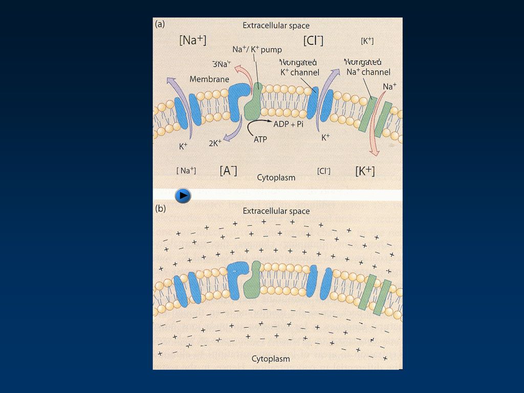
Due to differences in permeability of membrane to charged particles 전하를 이온, 유기물의 막 투과성 차이 음전하 유기물 비투과 completely impermeable to A- relatively permeable to K+ K+이온에 비교적 투과 relatively impermeable to Na+ Na+ 이온에 비교적 비투과 Inside of cell negative relative to the outside 세포 막 내 음전하 ~ -70 mV for many neurons At resting membrane potential, neither K+ nor Na+ are in equilibrium 지질 이중막 Fig 3.21 음전하 유기물 pump 막 투과

17
Electrical Activity of Neurons: Electrical Signals 신경세포의 전기적 활동: 전기적 신호
changes in membrane potential away from the resting membrane potential 정지 막전위로부터 膜電位의 변화 Depolarization 脫分極 decrease difference in charge btw ICF and ECF 막 내외의 電荷 차이 감소 Hyperpolarization 過분극 increase difference in charge btw intracellular and extracellular fluids 막 내외의 전하 차이 증가 Due to changes in membrane permeability and altering flow of charged particles 막 透過性과 전하입자의 이동 변화 Fig 4.9 기록전극 오실로스코프 탈분극/자극 과분극/억제

18
Membrane Proteins Involved in Electrical Signals
Non-gated (leak) ion channels “Always” open specific for a particular ion K+ Na+
 ion channels. Always open. specific for a particular ion. K+ Na+")
19
Membrane Proteins Involved in Electrical Signals 전기적 신호와 관련된 막단백질
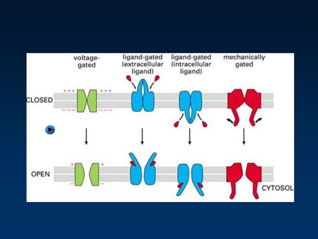
Gated ion channels 開閉性 이온통로 Open only in response to specific stimuli Major types in neurons Ligand-gated (-regulated) 리간드 依存 開閉 이온 通路 Binding of a chemical signal (ligand) causes channel to open 化學的 刺戟(例, 神經傳達物質)에 反應 Found on dendrites and cell body 세포체와 수상돌기에 많다
 리간드 依存 開閉 이온 通路. Binding of a chemical signal (ligand) causes channel to open. 化學的 刺戟(例, 神經傳達物質)에 反應. Found on dendrites and cell body. 세포체와 수상돌기에 많다.")
20
Membrane Proteins Involved in Electrical Signals
Voltage-gated (-regulated) 電壓依存 開閉 이온 通路 Changes in membrane potential (depolarization) cause channel to open 막전위의 변화(탈분극)에 의해 열림 Found in axons 軸索에 많다 Requires membrane to be depolarized to a certain degree from the resting potential (threshold membrane potential) 막전위가 易置까지 탈분극 If membrane depolarized to threshold, a cycle of activation (channel opening) followed by inactivation (closing) ensues 활동전위 생성 Fig 4.10 이온 通路 비활성화
 電壓依存 開閉 이온 通路. Changes in membrane potential (depolarization) cause channel to open. 막전위의 변화(탈분극)에 의해 열림. Found in axons 軸索에 많다. Requires membrane to be depolarized to a certain degree from the resting potential (threshold membrane potential) 막전위가 易置까지 탈분극. If membrane depolarized to threshold, a cycle of activation (channel opening) followed by inactivation (closing) ensues 활동전위 생성. Fig 이온 通路 비활성화.")
22
Membrane Proteins Involved in Electrical Signals
Na+/K+ pump Used to maintain electrochemical gradients for Na+ and K+ across the plasma membrane 막을 경계로 Na+와 K+의 전기화학적 구배를 유지함 Maintains resting potential 정지(안정,휴지) 막전위 유지 Active (require ATP) 에너지 소모 3 Na+ out, 2 K+ in / ATP
 막전위 유지. Active (require ATP) 에너지 소모. 3 Na+ out, 2 K+ in / ATP.")
23
Action Potentials Nerve Impulses 活動電位 神經衝擊
begins at the axon hillock, travels down entire length of axon 축색구로부터 축색을 따라서 전파 brief, rapid reversal of membrane potential 막전위의 잠시 빠른 역전 Large change (~ mV) 막전위의 큰 변화 Driven by the opening of voltage-gated Na+ and K+ channels 전압의존성 Na+ & K+ 이온통로의 열림에 의해 self-propagating - strength of signal maintained 자가 재생 long distance transmission 장거리 전파
 막전위의 큰 변화. Driven by the opening of voltage-gated Na+ and K+ channels. 전압의존성 Na+ & K+ 이온통로의 열림에 의해. self-propagating - strength of signal maintained 자가 재생. long distance transmission 장거리 전파.")
25
Types of Electric Signals: Action Potentials
"All or none" response 실무율 悉無律 axon hillock must be depolarized to the threshold potential for voltage-gated channels to open 전압의존성 통로를 열기 위해 축색구가 역치로 탈분극도어야 한다. if depolarized to (or beyond) threshold, voltage-gated channels will open and action potential (AP) will depolarize as much as possible 활동전위 생성 if threshold not reached, voltage-gated channels do not open and no AP will occur 역치하에서 활동전위 생성불능 Signal will quickly lose strength as it travels down axon 국소전위의 점진적 약화
 threshold, voltage-gated channels will open and action potential (AP) will depolarize as much as possible 활동전위 생성. if threshold not reached, voltage-gated channels do not open and no AP will occur 역치하에서 활동전위 생성불능. Signal will quickly lose strength as it travels down axon. 국소전위의 점진적 약화.")
26
Events During an Action Potential: Depolarization 탈분극
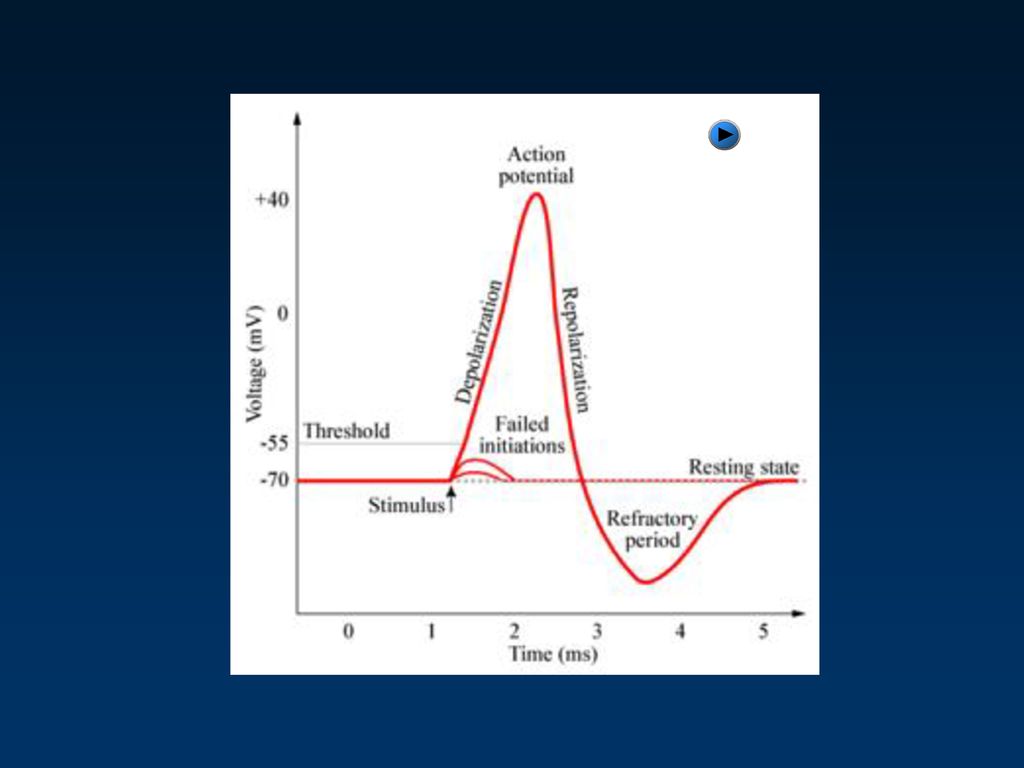
Triggering event (e.g., opening of ligand-gated channels) causes membrane to depolarize 리간드 의존 통로의 열림과 같은 촉발 사건에 의한 탈분극 Relatively slow increase until threshold potential is reached 역치에 느리게 다다름 Both voltage-gated Na+ channels (quickly) and K+ channels (slowly) begin to open 전압의존성 Na+통로가 빨리 열리고 K+통로는 늦게 열림 As voltage-gated Na+ channels open, Na+ enters cell 전압의존성 Na+통로가 열리면서 Na+이 유입 Positive feedback response 양성되먹임반응 further depolarization 더 탈분극 more channels open 더 통로 열림 Membrane rapidly depolarizes past 0 mV, reversing membrane polarity (to +30 mV) 0mV를 넘어 막 극성이 뒤바뀜 Depolarization is driven by the flow of Na+ into the cell
 causes membrane to depolarize. 리간드 의존 통로의 열림과 같은 촉발 사건에 의한 탈분극. Relatively slow increase until threshold potential is reached 역치에 느리게 다다름. Both voltage-gated Na+ channels (quickly) and K+ channels (slowly) begin to open. 전압의존성 Na+통로가 빨리 열리고 K+통로는 늦게 열림. As voltage-gated Na+ channels open, Na+ enters cell 전압의존성 Na+통로가 열리면서 Na+이 유입. Positive feedback response 양성되먹임반응. further depolarization 더 탈분극. more channels open 더 통로 열림. Membrane rapidly depolarizes past 0 mV, reversing membrane polarity (to +30 mV) 0mV를 넘어 막 극성이 뒤바뀜. Depolarization is driven by the flow of Na+ into the cell.")
27
Events During an Action Potential: Repolarization 재분극
When membrane potential reaches ~ +30 mV, Na+ channels are deactivated 소디움 통로 비활성화 No further flow of Na+ into the cell 소디움 이온 유입 불가 At same time, voltage-gated K+ channels open fully 포타슘 통로 완전히 열림 K+ flows out of the cell 포타슘 유출 membrane repolarizes toward resting potential 정지 막전위로 막의 재분극화 When membrane potential drops below threshold, K+ channels close 막전위가 역치 이하로 하강 시 포타슘 통로 닫힘 Na+ and K+ concentration gradients across the membrane restored by the Na+-K+ pump 펌프에 의한 Na+ 과 K+ 농도 구배의 회복 Repolarization is driven by the flow of K+ out of the cell

28
Action Potential Frequency and Stimulus Intensity 活動電位 頻度와 刺戟 强度
Response of the nerve cell to the stimulus is “all or none” 자극에 대한 신경세포의 반응은 실무율적 Amount of depolarization (amplitude) is always the same as long as depolarized to threshold 탈분극 정도는 역치에 이르면 항상 동일 differences in stimulus intensity are detected by the frequency of action potential generation 자극 강도는 활동전위의 빈도로 나타남 Stronger stimuli depolarize the membrane to threshold more quickly 강한 자극은 더 빨리 막을 탈분극시켜 역치에 이르게 함 APs are generated more frequently with increased stimulus intensity 자극강도 증가에 따라 더 많은 활동전위 발생
 is always the same as long as depolarized to threshold. 탈분극 정도는 역치에 이르면 항상 동일. differences in stimulus intensity are detected by the frequency of action potential generation. 자극 강도는 활동전위의 빈도로 나타남. Stronger stimuli depolarize the membrane to threshold more quickly. 강한 자극은 더 빨리 막을 탈분극시켜 역치에 이르게 함. APs are generated more frequently with increased stimulus intensity. 자극강도 증가에 따라 더 많은 활동전위 발생.")
29
Refractory Period 不應期 Fig 4.16
Interval that must pass before the neuron segment can undergo a second action potential 세포의 한 부위에서 두 번째 활동전위가 생성될 때 까지 기다려야 하는 시간간격 Membrane potential and ion concentration gradients must be restored 막 전위와 이온 농도구배의 회복 1. absolute refractory period 절대불응기 Neuron segment is in the process of undergoing an AP 활동전위생성기 Na+ channels are either fully open (depolarization) or in an inactive state (repolarization) Na+통로 완전 열린 상태(탈분극)나 비활성화 (재분극)상태 Cannot respond to a second stimulus 2. relative refractory period 상대적 불응기 Na+ channels can be activated again, but K+ still flowing out of the cell at a high rate Na+통로 재활성화 가능, K+ 多 유출 Action potential may be produced if a stronger stimulus is applied 强 자극에 의한 활동전위 발생가능 Fig 4.16
 or in an inactive state (repolarization) Na+통로 완전 열린 상태(탈분극)나 비활성화 (재분극)상태. Cannot respond to a second stimulus. 2. relative refractory period 상대적 불응기. Na+ channels can be activated again, but K+ still flowing out of the cell at a high rate Na+통로 재활성화 가능, K+ 多 유출. Action potential may be produced if a stronger stimulus is applied 强 자극에 의한 활동전위 발생가능. Fig")
30
Action Potential Propagation 활동전위의 전파
Action potentials are regenerated at each segment of the axon 축색 각 부위에서 활동전위의 재생 Na+ moving into one segment of the neuron during the depolarization phase quickly moves laterally inside the cell 탈분극 시 소디움 이온이 신경세포의 한 부분으로 유입되어 막내에서 빨리 측면 이동 Depolarizes adjacent segment to threshold, triggering an AP at the segment 옆 부분을 역치로 탈분극하여 활동전위 촉발 Action potentials are produced continuously along the plasma membrane of unmyelinated axons 무수축색의 막을 따라 활동전위가 계속적으로 발생 Fig 4.17

31
Action Potential Propagation: Myelinated Axons 활동전위의 전파: 유수축색
Myelination limits sites where an action potential occurs to the nodes of Ranvier 수초화에 따라 활동전위는 란비에 結節에서 발생 Na+ entering at node quickly passes to next node, bringing it to threshold 결절에 유입된 Na+은 다음 결절로 흘러가 역치로 막전위를 탈분극화 Saltatory conduction 도약전도 跳躍傳導 Action potential “jump” from one node to the next 결절에서 결절로 점프 AP conduction speed ( 50x) 전도속도傳導速度의 증가
 전도속도傳導速度의 증가.")
32
Synapses 神經連接 Synapse Communication junction between a neuron and either another neuron or a muscle or gland cell 소통접합: 신경세포간, 신경세포-근육(선세포) Chemical signals (neurotransmitters) released from axon terminal and bind to receptors on adjacent cell 축색종말에서 분비된 화학신호(신경전달물질)가 옆 세포의 수용체에 결합 Stimulates physiological change (usually change in membrane potential) in the recipient cell 수용세포의 생리적 변화 (막 전위의 변화)
 Chemical signals (neurotransmitters) released from axon terminal and bind to receptors on adjacent cell. 축색종말에서 분비된 화학신호(신경전달물질)가 옆 세포의 수용체에 결합. Stimulates physiological change (usually change in membrane potential) in the recipient cell. 수용세포의 생리적 변화 (막 전위의 변화)")
33
Anatomy of a Synapse presynaptic neuron synaptic cleft 연접간극(間隙)
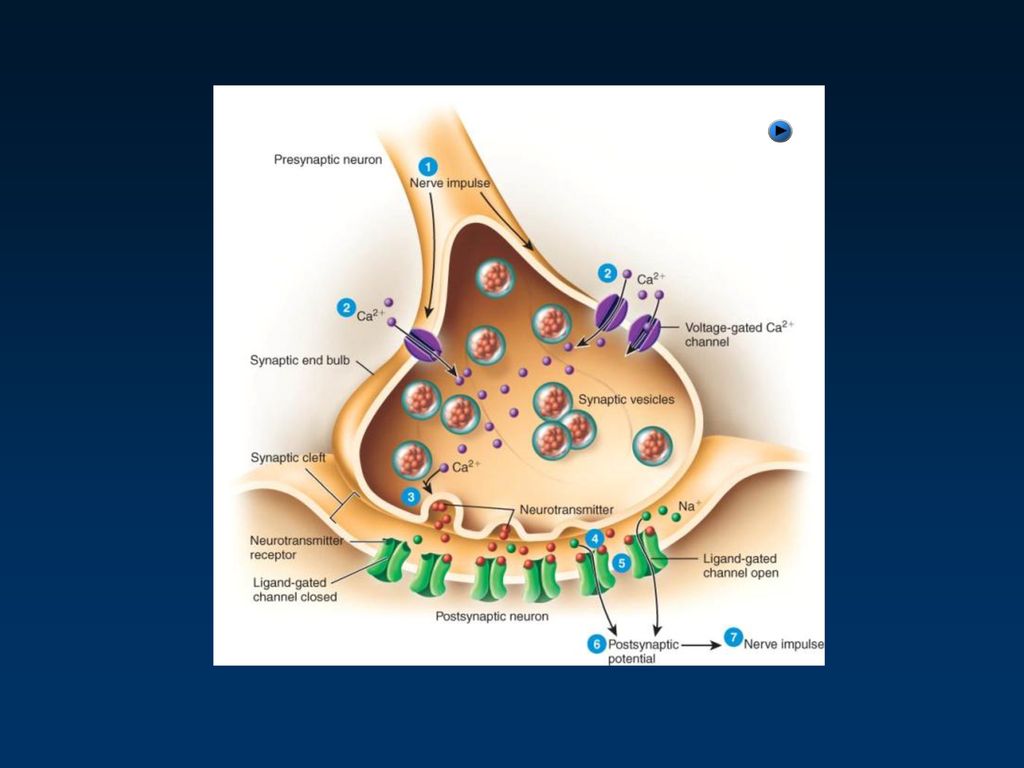
連接前細胞 連接後細胞 presynaptic neuron axon terminals contain synaptic vesicles filled with neurotransmitter synaptic cleft 연접간극(間隙) Narrow space between cells postsynaptic cell Contains receptor proteins that will bind neurotransmitter 受容器 단백질이 신경전달물질과 결합 Figs 4.19 and 4.20 連接前軸索終末 連接小囊 傳達物質 分泌 連接後細胞
 Narrow space between cells. postsynaptic cell. Contains receptor proteins that will bind neurotransmitter. 受容器 단백질이 신경전달물질과 결합. Figs 4.19 and 連接前軸索終末. 連接小囊. 傳達物質 分泌. 連接後細胞.")
34
連接後細胞의 軸索돌기 連接前細胞 末端 軸索돌기 軸索

35
Chemical Synapse Function
AP이 Ca2+ 통로 열리게 Ca2+ 유입 Ca2+ 에 의한 연접소낭의 exocytosis (吐細胞현상) 신경전달물질이 連接空間에서 확산하여 연접후 세포의 受容器에 결합 Fig 4.20
 신경전달물질이 連接空間에서 확산하여 연접후 세포의 受容器에 결합. Fig")
37
Chemical Synapse Function
연접후세포의 특정 리간드 의존성 이온통로의 열림 연접후세포의 막을 통해 이온 이동 연접후세포의 연접전위 유발 (흥분적/억제적) Figs
 Figs")
38
Chemical Synapse Function
연접전위의 2종류 1. Excitatory postsynaptic potential (EPSP) 흥분성 연접후 전위 Postsynaptic cell membrane depolarizes 2. Inhibitory postsynaptic potential (IPSP) 억제성 연접후 전위 Postsynaptic cell membrane hyperpolarizes EPSP가 충분히 탈분극하여 역치에 이르도록 강하면 → 연접후 세포에서 AP 발생
39
Characteristics of Synaptic Potentials
Decrease in amplitude with distance 국소공간적 감소 화학적 연접 부위로부터 멀어질수록 막전위가 감소한다. Graded responses 차등적 반응 신경전달물질이 많이 분비될수록 막전위가 크게 변함 Can summate 공간 및 시간적 加重 연접후 전위는 국소적이며 차등적, 비실무율적 시간적 공간적으로 가까운 EPSPs and IPSPs 의 합산효과 Aspects of summation Spatial summation – 여러 신경연접전 세포로 부터 신경연접 받음 Temporal summation – 신경연접전 세포로 부터 계속적인 신경전달물질 분비 Figs 4.21,
40
空間的 加重 時間的 加重

42
Examples of Neurotransmitters: Acetylcholine (ACh)
다양한 종류의 신경세포들에 있다. Ach 수용기에 따라 ESPSs or IPSPs E.g., Nicotinic ACh 수용기 특정 뇌영역, 골격근, 자율운동신경 Ach의 수용기 결합에 의해 이온통로 열림, 빠른 Na+ 유입, 느린 K+ 유출 EPSPs 생성 acetylcholinesterase 에 의한 Ach의 분해 수용기에 짧은 시간 동안 작용

43
Examples of Neurotransmitters: Acetylcholine (ACh)
E.g. Muscarinic ACh 수용기 CNS 신경세포, 평활근, 선세포, 심장근 수용기 (G-protein linked receptor) 는 이온통로와 독립적 ACh 결합에 의해 세포막의 G-proteins 활성화 G-protein 의 α and βγ subunits로 분해 곧 비활성화 (subunits재결합) G-proteins 에 의해서 세포 특이적인 효소와 통로의 활성화 E.g., 평활근에서 EPSPs E.g., 심장근에서 IPSPs Fig 4.28 and 4.29
 는 이온통로와 독립적. ACh 결합에 의해 세포막의 G-proteins 활성화. G-protein 의 α and βγ subunits로 분해. 곧 비활성화 (subunits재결합) G-proteins 에 의해서 세포 특이적인 효소와 통로의 활성화. E.g., 평활근에서 EPSPs. E.g., 심장근에서 IPSPs. Fig and")
44
Other Examples of Neurotransmitters / Receptors
GABA (gamma-aminobutyric acid) 주된 抑制性 傳達物質 Cl- 유입 과분극화 IPSPs 유발 Glutamate로 부터 合成 많은 GABA 作動藥: 安定劑 Valium and Xanax Picrotoxin, 拮抗劑, 痙攣 誘引劑 GABA 非活性化: 再吸收 Fig 4.27
 주된 抑制性 傳達物質. Cl- 유입 과분극화. IPSPs 유발. Glutamate로 부터 合成. 많은 GABA 作動藥: 安定劑. Valium and Xanax. Picrotoxin, 拮抗劑, 痙攣 誘引劑. GABA 非活性化: 再吸收. Fig")
45
Other Examples of Neurotransmitters
Monoamines Dopamine, norepinephrine, epinephrine (catecholamines) Serotonin Function through G-Protein linked receptors Leads to activation of enzymes and production of second messengers (e.g., cAMP) inside cell Second messengers activate additional enzymes (e.g., protein kinases), that induce metabolic changes or changes in membrane potential Monoamines generally taken back up by the pre-synaptic cell
 Serotonin. Function through G-Protein linked receptors. Leads to activation of enzymes and production of second messengers (e.g., cAMP) inside cell. Second messengers activate additional enzymes (e.g., protein kinases), that induce metabolic changes or changes in membrane potential. Monoamines generally taken back up by the pre-synaptic cell.")
46
Norepinephrine (noradrenalin) (NE)
중추 (시상하부, 뇌교의 청반) 및 말초 ANS 교감신경: 혈압상승 종말에서 합성 Adrenergic 수용기 subtypes, 수용기에 따라 + or - 기능 비활성화: 재 흡수 Cocaine, Methamphetamine 분비증가 및 재흡수 차단 Propanalol, an NE 길항제, 항고혈압제 (antihypertensive) Pseudoephedrine (Sudafed), Phenylpropanolamine (PPA) : NE 작동제
 및 말초. ANS 교감신경: 혈압상승. 종말에서 합성. Adrenergic 수용기 subtypes, 수용기에 따라 + or - 기능. 비활성화: 재 흡수. Cocaine, Methamphetamine 분비증가 및 재흡수 차단. Propanalol, an NE 길항제, 항고혈압제 (antihypertensive) Pseudoephedrine (Sudafed), Phenylpropanolamine (PPA) : NE 작동제.")
47
Dopamine (DA) Phenylalanine으로 부터 합성, 종말에서 NE의 先驅 物質
黑質(Substantia nigra) Cocaine, Methamphetamine :DA 분비 촉진 & 재흡수 차단 Parkinson’s Disease: 기저핵의 DA신경세포의 사멸 Haloperidol (Haldol) and clozapine (Clozaril): DA 길항제로 정신분열증 치료
 Cocaine, Methamphetamine :DA 분비 촉진 & 재흡수 차단. Parkinson’s Disease: 기저핵의 DA신경세포의 사멸. Haloperidol (Haldol) and clozapine (Clozaril): DA 길항제로 정신분열증 치료.")
48
Serotonine (5-HT) indoleamine 계열 봉선핵 (縫腺核 Raphe nuclei), 脊髓 後角
Tryptophan에서 합성 2%만 뇌 내, 수면, 식욕, 기분조절 비활성화: 재 흡수 LSD & mescaline: 작동제 어떤 5-HT 길항제: 구토 저하제로 불안, 우울증 치료제(재흡수 차단): Prozac, Celexa, Lexapro, Zoloft
: Prozac, Celexa, Lexapro, Zoloft.")
49
Peptide 신경 단백질 ~30개 amino acids, 지속, 장기적 효과
세포체 내 리보좀에서 합성->ER->Golgi체->분획->소낭->축삭이동->종말 Substance P: 통각정보 매운 고추의 생리활성 물질인 Capsaicin이 Substance P 분비 Endorphins and enkephalins: Opiate 기능, Substance P 분비 억제 Morphine and heroin: Substance P의 자연적 길항제 달리기 선수, 임산부의 출산 Vicodin (hydrocodone) and Percocet (oxycodone) and other Opioids: Substance P의 인공 길항제
 and Percocet (oxycodone) and other. Opioids: Substance P의 인공 길항제.")
Similar presentations

. Why must organisms reproduce?>")
 2009103850 류문영, 2006200408 마진영, 2009103851 박주원 4 TEAM.>")


. 생명의 특징 (attributes of living matter) Six most important life processes Metabolism ( 신진대사 ) Responsiveness ( 반응, adaptation.>")


