Download presentation
Presentation is loading. Please wait.

1
Molecular Imaging of Cancer
Jae Min Jeong, Ph.D. Department of Nuclear Medicine Seoul National University College of Medicine

2
분자 영상법 정의 : 세포 또는 조직내의 분자유전학적 변화를 특정 유전자를 이용하여 영상화하는 첨단기법 종류 : 적용 범위 :
세포 또는 조직내의 분자유전학적 변화를 특정 유전자를 이용하여 영상화하는 첨단기법 종류 : 1. 광학 영상법 : 세포, 소동물 수준 (Luciferase, Fluorescence Protein) 2. 핵의학 영상법 : 세포, 소동물, 사람 수준 (Sodium Iodide Symporter, HSV1-tk, D2R) 3. MR 영상법 : 대동물, 사람 수준 적용 범위 : 1. 내인성 또는 외인성 유전자 발현 2. 단백질 간의 상호작용 3. 신호전달 체계 4. 생체 내 세포추적
 2. 핵의학 영상법 : 세포, 소동물, 사람 수준. (Sodium Iodide Symporter, HSV1-tk, D2R) 3. MR 영상법 : 대동물, 사람 수준. 적용 범위 : 1. 내인성 또는 외인성 유전자 발현. 2. 단백질 간의 상호작용. 3. 신호전달 체계. 4. 생체 내 세포추적.")
3
분자 생물학적 평가 vs 분자 영상 기법 기존의 연구방법 분자영상 기법 (분자 생물학적 방법) 생체 조직 한 개체의 반복
필요 (대상물의 희생이나 손상) 불가능 많은 수 필요 많다 불필요 (개체 자체를 영상화) 가능 적은 수 필요 적다 기존의 연구방법 분자영상 기법 (분자 생물학적 방법) 생체 조직 한 개체의 반복 대상물 시간 소요 생체조직을 손상하지 않고(Non-invasive) 반복적으로 (Repeatable) 정량화(Quantification)가능
 불가능. 많은 수 필요. 많다. 불필요. (개체 자체를 영상화) 가능. 적은 수 필요. 적다. 기존의 연구방법 분자영상 기법. (분자 생물학적 방법) 생체 조직. 한 개체의 반복. 대상물. 시간 소요. 생체조직을 손상하지 않고(Non-invasive) 반복적으로 (Repeatable) 정량화(Quantification)가능.")
4
Monitoring reporter gene expression
Reporter gene combination with target gene - Express same mRNA transcript Classic : -gal, AP, luciferase, fluorescence protein, NIS, Tk

5
Optical Images 1. Fluorescence excitation on external light source
2. Luminescence chemically powered light source luciferin and luciferase

6
Why optical image? 1. Powerful labeling technique gene expression
2. Easy quantitation amount of light vs. number of cells 3. Extremely low background 4. Relatively simple instruments

7
IVIS Imaging System Control computer Camera controller Imaging chamber
Cryogenic refrigerator

8
Nature Genetics, 4: 613 (2003), Dr. Fraser
, Dr. Fraser")
9
The novel candidates for MI
Global Views of Human Cancer Gene Expression Imaging of Antibodies and Phage Targeted to Blood Vessels of Tumors Bioactive Ligands for Peptide Receptors Reverse Engineering of Targeting Ligands Based on Expressed Epitope Patterns

10
Use of TG mouse expressing fluorescent protein in mammalian development and neurobiology
Nature Genetics, 4: 613 (2003), Dr. Fraser
, Dr. Fraser.")
11
Combinatorial fluorescent protein
Reporter detection in live chimera And double transgenics A-c: embryo, cyan : epiblast Yellow: endoderm and trophoblast D-f: chimeric adult organ f: liver: greater cell intermingling during development G: neurons in brain Thy-1 double TG mouse Nature Genetics, 4: 613 (2003), Dr. Fraser
, Dr. Fraser.")
12
1. To follow movement of cells in living mouse embryo
-TG mouse with EGFP under Oct4 promoter 2. Developing kidney -TG mouse with HoxB7-EGFP 3. Blood Flow dynamics in live embryo - With EGFP under e(embryonic)-globin promoter Nature Genetics, 4: 613 (2003), Dr. Fraser
-globin promoter. Nature Genetics, 4: 613 (2003), Dr. Fraser.")
13
Fluorescent fusion protein as Markers of subcellular compartments
Nature Genetics, 4: 613 (2003), Dr. Fraser
, Dr. Fraser.")
14
Imaging of Antibodies and Phage Targeted to Blood Vessels of Tumors
Trends in Mol Med, 8: 563, 2002 Using in vivo phage display, they can identify the organ-specific and disease-specific protein expressed on endothelial surface of blood vessel

15
Cell Migrations: Two Photon Imaging of Lymphocyte Motility and Antigen presentation in Vivo PNAS, 100: 2604, 2003 Science, 296:1869, 2002 Two photon laser microscopy naïve T cell motility and migration in lymph nodes

17
Science 296: 1869, 2002 T=25 um/min B= 25/2 um/min

18
Luciferase를 이용한 광학적 영상법
반딧불이에서 유래 기질인 luciferin을 oxyluciferin으로 변화시 나오는 에너지를 빛 으로 반응 광학 기술의 발달로 개체 수준으로 측정 가능 : LAS-1000 (fuji)
")
19
Reporter gene imaging for signal transduction pathway of
G-protein coupled receptor: D2 receptor Dopamine2 receptor signal pathway: D2 receptor biding Dopamine -> activation of a Gi-protein complex -> inactivation of adenylyl cyclase -> decrease in cellular cAMP level -> cAMP activate PKA (protein kinase A) - - - > CREB (cAMP response element-binding protein: transcriptinal factor) phospholylate by PKA -> CRE controlled gene exp. Vector pCMV-CREB pG5-FLUC pCMV-D2R pCMV GAL4 CREB GAL4 BS Luci D2R
 > CREB (cAMP response element-binding protein: transcriptinal. factor) phospholylate by PKA -> CRE controlled gene exp. Vector. pCMV-CREB. pG5-FLUC. pCMV-D2R. pCMV. GAL4. CREB. GAL4 BS. Luci. D2R.")
20
In vitro, Group 1 -> pCMV-CREB and pG5-FLUC transiently transfect to 293 T cell Group 2 -> three plasmids transfect To treatment of different conc. of dopamine (0-200uM), and then Luciferase assay In vivo, Group 1 -> pCMV-CREB and pG5-FLUC transfected cells (1x10⁶) were s.c. injected nude mice Group 2 -> three plasmids transfected cells were s.c. injected nude mice L-DOPA were i.f. injected nude mice
, and then Luciferase assay. In vivo, Group 1. -> pCMV-CREB and pG5-FLUC transfected cells (1x10⁶) were s.c. injected nude mice. Group 2. -> three plasmids transfected cells were s.c. injected nude mice. L-DOPA were i.f. injected nude mice.")
21
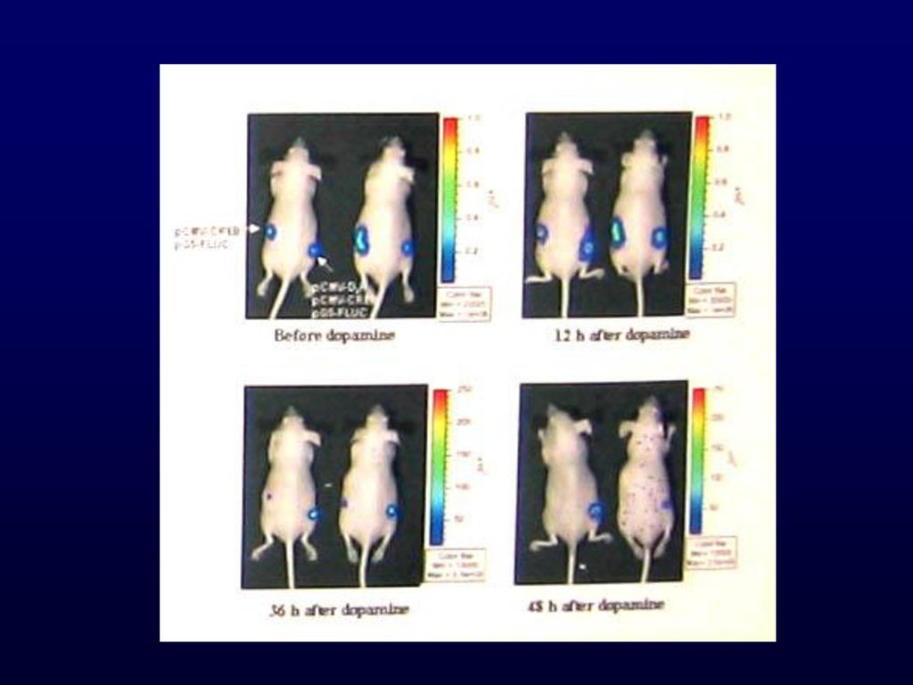
Result (in animal study)
Before dopamine -> group 1 and 2 cells showed strong bioluminescence signal 12h after dopamine -> group 2 cells significantly increased 36h after dopamine -> group 1 cells disappeared signal group 2 cells still showed signal

23
Modulation of a split synthetic renilla luciferase
complementation-based protein-protein interaction using the small molecule rapamycin in cells in living animals The mammalian target of rapamycin (mTOR) is a Ser/Thr protein kinase that functions in a signaling pathway lead to cell growth and/or proliferation. Rapamycin binds with the FRB with a molecular weight of 12000(FKBP12). FRB: FKBP12-rapamycin binding domain mTOR lacking the FRB is not capable of supporting G1 phase.
 is a Ser/Thr protein kinase. that functions in a signaling pathway lead to cell growth and/or. proliferation. Rapamycin binds with the FRB with a molecular weight of 12000(FKBP12). FRB: FKBP12-rapamycin binding domain. mTOR lacking the FRB is not capable of supporting G1 phase.")
24
Two vectors transfect to 293 T cell.
pCMV N-Luci FRB pCMV FKBP12 C-Luci Two vectors transfect to 293 T cell.

25
The cells exposed to 10nM rapamycin
for 24hrs showed significant increase in hrluc activity.

26
Luciferase를 이용한 광학적 영상법
LAS 1000 Luciferase를 이용한 광학적 영상법 Construction of SK-HEP1-NIS-Luc SK-HEP1 (human hepatocarcinoma) NIS-Luc : IRES sequence (pIRES)
 NIS-Luc : IRES sequence (pIRES)")
27
LAS 3000 RA처리후 48시간 영상 등 RA no treat RA 2mg/20g RA 4mg/20g
SK-HEP1 (8ⅹ106) SK-RARE (1ⅹ106) SK-RARE (8ⅹ106) SK-RARE (4ⅹ106) 등 RA no treat RA 2mg/20g RA 4mg/20g
 SK-RARE (1ⅹ106) SK-RARE (8ⅹ106) SK-RARE (4ⅹ106) 등. RA no treat. RA 2mg/20g. RA 4mg/20g.")
28
Radio reporter genes Visualize opaque tissue
Quantify expression level of reporter gene Noninvasive, repetitive Advantages of radio nuclide-based methods Highly sensitive : 10-12mol/L of radio substrate good for weak promoter Highly quantitative : dynamic image, kinetic model

29
Sodium/Iodide Symporter (NIS)
분포: 갑상선 세포에서 주로 발현 기능: 갑상선 세포 내로의 요오드섭취에 중요한 역할 리포터 유전자로서의 장점 비싼 PET 스캐너, 복잡한 방사성 의약품이 필요 없음 방사성요오드로 쉽게 gamma-camera로 영상획득 가능 요오드의 생체 내에서의 대사과정이 잘 알려져 있음 요오드가 세포내 다른 분자들과 상호작용 하지않으므로 안전함

30
Sodium/iodide symporter (NIS)
(Dai G, et al. Nature 1996) Rat NIS (rNIS) protein : 618 amino acids Human NIS (hNIS) protein : 643 amino acids 84% identical to rNIS Structure : 13 transmembrane domains + 3 extracellular glycosylation sites 30 Seoul National University Hospital
 Rat NIS (rNIS) protein : 618 amino acids. Human NIS (hNIS) protein. : 643 amino acids. 84% identical to rNIS. Structure. : 13 transmembrane. domains + 3 extracellular. glycosylation sites. 30. Seoul National University Hospital.")
31
N K + Sodium/Iodide Symporter (NIS) : Na+ I- Na+ 분포: 갑상선 세포에서 주로 발현
분포: 갑상선 세포에서 주로 발현 기능: 갑상선 세포 내로의 요오드섭취에 중요한 역할 Na+ I- N K + Na+ 31 Seoul National University Hospital

32
Delivery of NIS gene into target cells
: 131I or 188Re administration (Radionuclide Gene Therapy) : 123I or 99mTc administration (Imaging)
 : 123I or 99mTc administration (Imaging)")
33
암세포에 NIS유전자 이입후 방사성 요오드 섭취율
Time (min) 30 60 90 120 Iodide uptake (pmol/106 cells) 100 200 300 400 500 600 ARO ARO-NIS ARO-NIS + KClO4
 Iodide uptake (pmol/106 cells) ARO. ARO-NIS. ARO-NIS + KClO4.")
34
NIS유전자 이입 암세포에서의 핵의학 영상 II
Tc-99m Re-188

35
NIS유전자 이입 암세포에서의 핵의학 영상 A B A: HSV1-tk 영상 (UCLA) B: NIS 영상 (본 연구실)
 B: NIS 영상 (본 연구실)")
36
PET reporter gene By receptor : bond radio ligand probe
Dopamine 2 Receptor By enzyme : sequester radio substrate probe - Herpes Simplex Virus 1 Thymidine Kinase (HSV1-tk)
")
37
Imaging the Expression of a Reporter Gene
Vector for Reporter Gene Delivery Reporter Gene HSV1-tk Radiolabeled Probe Promoter/Enhancer Constitutive Inducible [*I]-FIAU TK DPK TPK [*I]-FIAU [*I]-FIAU-(PO4) HSV1-TK mRNA HSV1-tk Enzyme HSV1-TK
 HSV1-TK. mRNA. HSV1-tk. Enzyme. HSV1-TK.")
38
The Paradigm of TKGFP Reporter Gene Imaging.
PET Image FIAU N H O F I FIAU-MP N H O P O4 F I FIAU N H O F I ATP TKeGFP

39
HSV1-tk As a reporter gene Thymidine derivative reporter probes
FIAU, IVDU(Iodovinyl-deoxyuridine) Used for both PET, SPECT Guanosine derivative reporter probes GCV, PCV, FHBG For PET
 Used for both PET, SPECT. Guanosine derivative reporter probes. GCV, PCV, FHBG. For PET.")
40
Improvement Strategies Better Sensitivity & Specificity
Improve the gene Novel approach - molecular biology Improve the probe Traditional approach - chemistry

41
Improve the Gene HSV1-tk -> HSV1-sr39tk ATC TTC CTC TTC ATG I F L
Base Position 811 HSV1-tk CTC ATC TTC GCC CTC Amino Acid L I F A L HSV1-sr39tk ATC TTC CTC TTC ATG I F L F M Gambhir et al., PNAS 97:2785,2000

42
Improve the Gene HSV1-tk -> HSV1-sr39tk
Gambhir et al., PNAS 97:2785,2000

43
Improve the Probe Pyrimidine Nucleosides Acycloguanosines
Probe R1 R2 R3 FIAU I F H FIRU I H F FMAU CH3 F H IVFRU CH2=CH2-I H F IUdR I H H BrUdR Br H H Pyrimidine Nucleosides HN H2N GCV O OH H PCV CH2 OH H FGCV O OH F FPCV CH2 OH F FHPG O F H FHBG CH2 F H Acycloguanosines

44
Paired FIAU - FHBG Comparison
Coronal Images Axial Images % dose/g % dose/g 0. 20+ 1.4+ 0. 20+ 1.4+ RG2 RG2TK+ RG2 RG2TK+ RG2TK+ RG2 RG2TK+ RG2 0.0 0.0 0.0 0.0 FHBG FIAU FHBG FIAU @ 2hr 12/13/00 @ 2hr 12/14/00 @ 2hr 12/13/00 @ 2hr 12/14/00 Tjuvajev et al. J Nucl Med, 2002 (in press)
")
45
Paired FIAU - FHBG Comparison
[18F]-FHBG Time (min) [124I]-FIAU % Dose/cc TdR (ml/g)) FIAU or FHBG (ml/g) RG2TK+ Cells Tjuvajev et al. J Nucl Med, 2002 (in press)
 [124I]-FIAU. % Dose/cc. TdR (ml/g)) FIAU or FHBG (ml/g) RG2TK+ Cells. Tjuvajev et al. J Nucl Med, 2002 (in press)")
46
Feb. 2003 Cell tracking, molecular targets, drug discovery
Ralph Weissleder Harvard Medical School Clinical oncology, imaging drug targets Patricia Price U. Manchester Protein interactions, imaging tools David Piwnica-Worms Washington U. Tracking of gene therapy, gene activities Sam Gambhir U. California, Los Angeles Imaging of gene expression Ronald Blasberg Memorial Sloan-Kettering Cancer Center PROJECT RESEARCHER Others in MOLECULAR IMAGING Feb. 2003
![]()
Similar presentations


 개발 장 규 태 참여기업: 대한바이오㈜ 한국생명공학연구원.>")

 –Leptin: apheliotrophic actions Atherosclerosis,>")


>")

법과>")
>")
