Download presentation
Presentation is loading. Please wait.

1
망막 컨퍼런스 AP. 박영훈/R3 정병주

2
Case 1

3
Chief complaints F/70 전ㅇ숙 Decreased VA & blurred vision
상기 환자 평소 침침함 있어 local 안과 방문하여,안저검사상 좌안 시신경 주위 망막 염증 및 부종 소견 보여 큰병원 권유받아 내원

4
Past History DM/HBP (-/-) Ocular op/trauma(-/+)
3년 전 cat.op(OU) at local Gls (-) Eye drop (-) F.Hx n-s
 at local. Gls (-) Eye drop (-) F.Hx n-s.")
5
Ocular examination VA OD 0.63(1.0) OS 0.8 IOP OD 10 mmHg OS 10 mmHg
Cornea : clear AC : deep & cell(-) Pupil : round & nl. sized Lens : PCL in situ
 Pupil : round & nl. sized. Lens : PCL in situ.")
6
Ocular examination Fundus exam OD: nl. Optic disc c flat post pole
OS : suspiscious pre- & para optic disc membrane or optic disc drusen vit. Cell(trace)
")
7
FAG 10-15 sec 동맥기( 13) 15-20 동정맥기(laminar flow) 20-25 정맥기
7분 – 후기 : leaking 지속적으로 보임

8
Ocular examination

9
Initial Laboratory exam

10
Initial Laboratory exam

11
Initial Laboratory exam

12
Initial Laboratory exam
CHEST PA diffuse micronodular opacities in the both lungs Diffuse interstial lung disease is suggested

13
Cue list & Initial plan Impression Plan
Fd & FAG : retinal vasculitis 소견 Lab : elevated c-ANCA & ACE, ANA(+) Chest PA : diffuse ILD Impression R/O retinal vasculitis d/t rheumotologic origin(OS) Plan Rheumatology consult O-LON X 4, O-LVF X 4 (OS)
 Chest PA : diffuse ILD. Impression. R/O retinal vasculitis d/t rheumotologic origin(OS) Plan. Rheumatology consult. O-LON X 4, O-LVF X 4 (OS)")
14
Progress note Final diagnosis Sarcoidosis with ILD Ocular sarcoidosis
HRCT Diffuse lung infiltration Pulmonology consult CS 에서 VATS Bx.시행 Final diagnosis Sarcoidosis with ILD Ocular sarcoidosis Conjunctival & corneal lesion(-) Iris nodule(-) Ant. Uveitis(-) retinal vasculitis c NV at disc(+) Chest PA : abnormal ACE : elevated :: VATS biopsy ::
 Iris nodule(-) Ant. Uveitis(-) retinal vasculitis c NV at disc(+) Chest PA : abnormal. ACE : elevated. :: VATS biopsy ::")
15
Case 2

16
Chief complaints F/66 김ㅇ자 Decreased VA(OD>OS) Onset ) 2months ago
2 개 월 전부터 양안이 뿌연 증상 있어(우>좌), 강릉 아산병원 방문 양안 포도막염 진단하에 안약 점안하며 치료하던 중 (류마티스 내과 진료도 보았으나, 확실히 진단받은 것은 없음), 본원 5 일 전부터 우안 상측 시야 장애 있어 3 일전 FAG 시행 후 BRAO(OD) 진단받고 further evaluation 위해 내원
, 강릉 아산병원 방문 양안 포도막염 진단하에 안약 점안하며 치료하던 중 (류마티스 내과 진료도 보았으나, 확실히 진단받은 것은 없음), 본원 5 일 전부터 우안 상측 시야 장애 있어 3 일전 FAG 시행 후 BRAO(OD) 진단받고 further evaluation 위해 내원.")
17
Past History DM/HBP (-/+) for 3yrs, PO medi Ocular op/trauma(-/-)
강릉아산병원 류마티스 내과에서 H-LON 40mg Hydroxychloroquine 복용 중 Ocular op/trauma(-/-) Gls (-) Eye drop (+) O-COST X 4 O-BMDP X 4 O-MOX X 4 /OU F.Hx n-s
 Gls (-) Eye drop (+) O-COST X 4. O-BMDP X 4. O-MOX X 4 /OU. F.Hx n-s.")
18
Ocular examination VA OD 0.25(N-C) OS 0.5(N-C) IOP 17/15 mmHg
Cornea : clear AC : deep & cell(trace) Pupil : round & normal sized Lens : moderate cortical opacity / OU
 Pupil : round & normal sized. Lens : moderate cortical opacity / OU.")
19
Ocular examination Fundus exam
OU : nl. Optic disc c thin ERM , multiple infiltration at mid pph. Vitreous cell/opacity(++) OD : R/O BRAO
 OD : R/O BRAO.")
20
FAG 15 sec 20 sec 맥락막 순환 – 동맥기(12-15 sec) – 동정맥기 – 정맥기(20-25 sec) – 재순환기 – 후기 BRAO – 불투명한 망막이 명확히 구분, 동맥기에 충만이 전혀 되지 않음. 허혈성 망막의 저형광 소견

21
FAG 후기의 미만성의 과형광 소견

22
Ocular examination

23
Ocular examination

24
Initial Laboratory exam

25
Initial Laboratory exam

26
Initial Laboratory exam

27
Initial Laboratory exam
CHEST PA no evidence of definite abnormality except for mild cardiomegaly.

28
Cue list 2 개월 전 부터 양안 포도막염 증상
Local clinic 에서 Rheumatic ds. 로 H-LON, hydroxychlroquine 복용 Fd : multiple infiltration at mid pph. vitreous cell/opacity(++) FAG : R/O BRAO(OD) Lab : ANA(+) Lab : Toxocariosis Ig G(+)
 FAG : R/O BRAO(OD) Lab : ANA(+) Lab : Toxocariosis Ig G(+)")
29
Impression & Plan Impression Plan
R/O occlusive vasculitis d/t unknown origin(OU) Plan Rheumatology, oncology, infection, cardiology, hematology consult O-LON X 4, O-DCF X 4, O-LVF X 4, O-COST X 2 /OU H-LON tapering + implanta 200mg Diagnostic ppV 고려
 Plan. Rheumatology, oncology, infection, cardiology, hematology consult. O-LON X 4, O-DCF X 4, O-LVF X 4, O-COST X 2 /OU. H-LON tapering + implanta 200mg. Diagnostic ppV 고려.")
30
Systemic Workup Rheumatology : N-S Oncology : N-S
FANA 1:00 및 rheumatic lab 결과상(-) Oncology : N-S abdominal CT, duodenoscopy, colonoscopy, tumor marker Cardiology : both hilar LAP CAD 발견 및 treatment start Infection : N-S Toxocariasis 및 기타 viral marker 에서 이상소견(-) Hematology chest CT(enhance, lung mediastinum), abdomen pelvis CT (enhance), neck CT( enhance ), CSF study, Brain MRI
 Oncology : N-S. abdominal CT, duodenoscopy, colonoscopy, tumor marker. Cardiology : both hilar LAP. CAD 발견 및 treatment start. Infection : N-S. Toxocariasis 및 기타 viral marker 에서 이상소견(-) Hematology. chest CT(enhance, lung mediastinum), abdomen pelvis CT (enhance), neck CT( enhance ), CSF study, Brain MRI.")
31
Operation record Op name
phaco+ppV+ PCL +endolaser +FAE + IOL insertion (OD) Cytology, microbial PCR(VZV, Toxoplasma DAN, HSV type 1, CMV), Gram stain & microbial culture -> non specific findings
 Cytology, microbial PCR(VZV, Toxoplasma DAN, HSV type 1, CMV), Gram stain & microbial culture. -> non specific findings.")
32
Progress note Hematology F/U B2 microglobulin
chest CT에서 mild enlarged Hilar & mediastinal LN, mild ILD 소견 Lymphoma 배제할 수 없어 PET CT 시행하기로 함. PET CT : lymphoproliferative or granulomatous ds. TB 및 sarcoidosis 가능성에 대해 pulmonology & CS F/U 하기로 함.(ACE : WNL) VATS biopsy 시행하기로 함
 VATS biopsy 시행하기로 함.")
33
Progress note Pathology Final diagnosis Sarcoidosis with ILD
Chronic granulomatous inflammation, consistent with Sarcoidosis. Final diagnosis Sarcoidosis with ILD Ocular sarcoidosis Conjunctival & corneal lesion(-) Iris nodule(-) Ant. Uveitis(+) Vitritis(+) Occlusive retinal vasculitis(+) Chest PA : WNL ACE : WNL :: VATS biopsy ::
 Iris nodule(-) Ant. Uveitis(+) Vitritis(+) Occlusive retinal vasculitis(+) Chest PA : WNL. ACE : WNL. :: VATS biopsy ::")
34
Progress note POD # 1month VA : 0.06/0.25 IOP : 12/16 mmHg
AC : deep & cell(-) Fd : flat(OD) stationary infiltration (OS) Plan : H-LON 10mg + Implanta 100mg O-LON X 4, O-MOX X 4, O-COST X 2 /OU Pulmonology 와 협진하에 medication 조절 예정 Subtenon triamcinolon inj.(OD) 시행함
 Fd : flat(OD) stationary infiltration (OS) Plan : H-LON 10mg + Implanta 100mg. O-LON X 4, O-MOX X 4, O-COST X 2 /OU. Pulmonology 와 협진하에 medication 조절 예정. Subtenon triamcinolon inj.(OD) 시행함.")
35
Case 3

36
Chief complaints M/39 양ㅇ호 Decreased VA & ocular pain(OD)
Onset ) 좌안은 어릴 때 부터 광각무상태, 우안은 일상생활 가능한 정도로 지냄 2010년 가을경 부터 간헐적으로 암시 증상이 발생하였음 경부터 갑자기 우안 통증 및 시력저하 증상 있어 local clinic 에서 ACG 의심하에 mannitolization 및 안압 하강제 점안 후 증상 완화되었음 울산대병원 내원하여 당시 시력은 HM, 안압 6mmHg, B scan 상 망막 박리 의심되어 본원 내원함.
 좌안은 어릴 때 부터 광각무상태, 우안은 일상생활 가능한 정도로 지냄. 2010년 가을경 부터 간헐적으로 암시 증상이 발생하였음 경부터 갑자기 우안 통증 및 시력저하 증상 있어 local clinic 에서 ACG 의심하에 mannitolization 및 안압 하강제 점안 후 증상 완화되었음 울산대병원 내원하여 당시 시력은 HM, 안압 6mmHg, B scan 상 망막 박리 의심되어 본원 내원함.")
37
Past History DM/HBP (-/-) Ocular op/trauma(-/-) Gls (-)
Prematurity는 아닌 것으로 알고 있음 Ocular op/trauma(-/-) Gls (-) Eye drop (+) : 내원 당시 안압하강제(-) O-LON X 4 CRAVIT X 4 O-2HOM X 4 /OD F.Hx n-s
 Gls (-) Eye drop (+) : 내원 당시 안압하강제(-) O-LON X 4. CRAVIT X 4. O-2HOM X 4 /OD. F.Hx n-s.")
38
Ocular examination VA OD HM(N-C) OS LP(-)
IOP OD 45mmHg (산동전) -> 70 mmHg (산동후) OS <5 mmHg Cornea : mild edematous c diffuse microbullae AC 1.5 at CAC, <1/4 at PAC & cell(-) Pupil : round & moderate dilatated state Lens : intumescent cataract / OD Nystagmus(+)
 -> 70 mmHg (산동후) OS <5 mmHg. Cornea : mild edematous c diffuse microbullae. AC 1.5 at CAC, <1/4 at PAC & cell(-) Pupil : round & moderate dilatated state. Lens : intumescent cataract / OD. Nystagmus(+)")
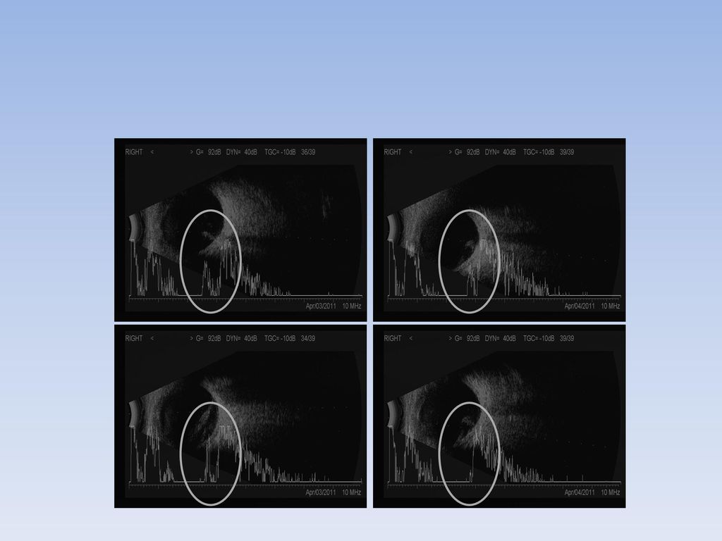
40
OD OS

41
Cue list 어릴 때 부터 시력 불량, 좌안은 실명 상태 AC : shallow anterior chamber depth
Lens : mature cataract Microphthalmia B scan : R/O RD

42
Impression & Plan Impression Plan R/O RD(OD) R/O lens induced glaucoma
Phaco+ppV under general anesthesia

43
Operation record Op name Postoperative diagnosis
ppV+ppL+MP+FAE+PFCL inj.+PFCL removal+endolaser+S.encircling+SO inj.(OD) Postoperative diagnosis PHPV c Total RD c PVR(OD)
 Postoperative diagnosis. PHPV c Total RD c PVR(OD)")
45
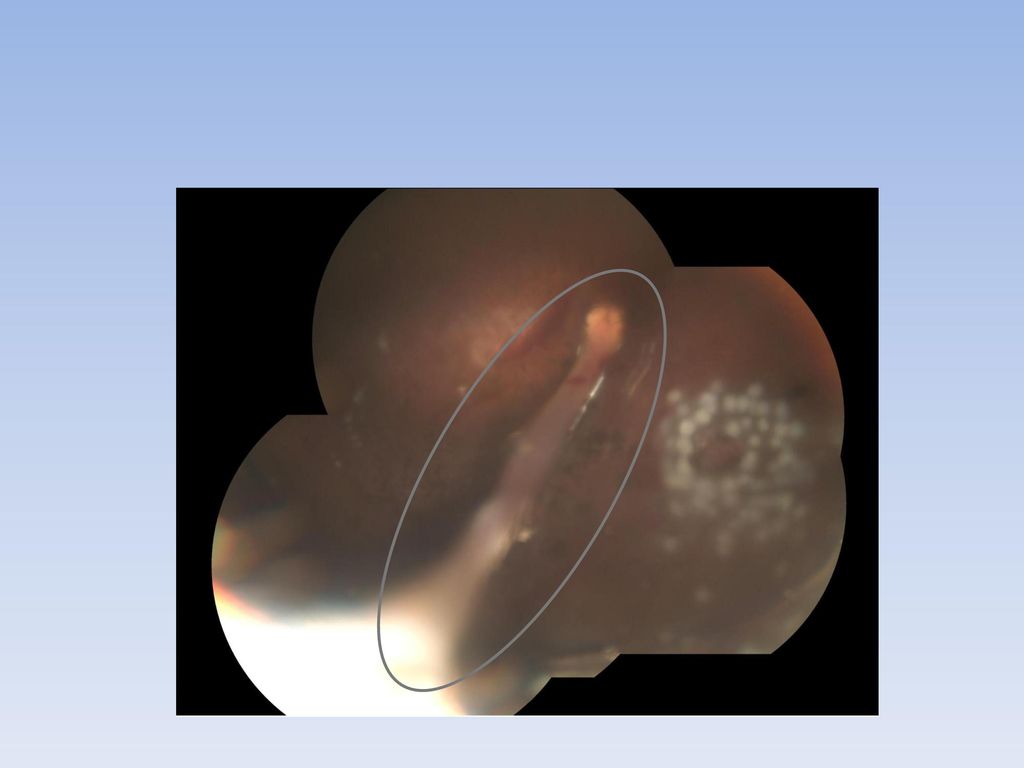
Progress note POD #1 VA : LP(+)
IOP(AP) 30 /O-COST x 2, O-BMDP x 2, O-TVT x hs Cornea : mild edematous c 3x4 mm sized epiedefect AC : 2.5 & cell(+++, RBC) Lens : A. aphakia Fd : Gliserting app. d/t SO, stalk arising from disc, seems to be flat post.pole, well-applied laser scars at 5 o/c, well elevated encircling band / OD
 30 /O-COST x 2, O-BMDP x 2, O-TVT x hs. Cornea : mild edematous c 3x4 mm sized epiedefect. AC : 2.5 & cell(+++, RBC) Lens : A. aphakia. Fd : Gliserting app. d/t SO, stalk arising from disc, seems to be flat post.pole, well-applied laser scars at 5 o/c, well elevated encircling band / OD.")
46
Progress note POD #3 VA : HM(+)
IOP(AP) 14 /O-COST x 2, O-BMDP x 2, O-TVT x hs Cornea : mild edematous c nearly healed epidefect AC : 2.5CT at center & cell(+, RBC) Lens : A. aphakia Fd : Gliserting app. d/t SO, stalk arising from disc, seems to be flat post.pole, well-applied laser scars at 5 o/c, well elevated encircling band / OD
 14 /O-COST x 2, O-BMDP x 2, O-TVT x hs. Cornea : mild edematous c nearly healed epidefect. AC : 2.5CT at center & cell(+, RBC) Lens : A. aphakia. Fd : Gliserting app. d/t SO, stalk arising from disc, seems to be flat post.pole, well-applied laser scars at 5 o/c, well elevated encircling band / OD.")
Similar presentations


 recent 2wks ago remote 10yrs ago Present Illness 72/M, HTN, angina 로 본원 IC(Pf. 김우식 ), DM 으로 본원 IE(Pf. 오승준 ) f/u 하는.>")
 Adm 2012.03.28 주소 Cough onset time) 내원 1 달 전 현병력 60 세 여자환자. HTN, dyslipidemia 로 타 병원에서 medication.>")


>")

