Download presentation
Presentation is loading. Please wait.

1
KCP 783 이대목동병원 박희정

2
건강검진상 시행한 흉부 컴퓨터 단층 촬영(CT)에서 결절이 발견
55세 남자 건강검진상 시행한 흉부 컴퓨터 단층 촬영(CT)에서 결절이 발견 기관지 내시경상에서 혈관이 풍부한 기관지내 종괴가 관찰되었습니다. 기관지 내시경 검사 중 종괴에서 세침흡인을 시행하였고 보내드린 슬라이드는 세침흡인 세포 도말입니다.
에서 결절이 발견. 기관지 내시경상에서 혈관이 풍부한 기관지내 종괴가 관찰되었습니다. 기관지 내시경 검사 중 종괴에서 세침흡인을 시행하였고 보내드린 슬라이드는 세침흡인 세포 도말입니다.")
3
흉부 컴퓨터 단층 촬영(CT)에서 우중엽의 기관지 내강을 채우는 조영증강이 잘되는 약 3cm 크기의 종괴가 관찰되었고 주변에 조영증강이 잘 되는 크기가 커진 림프절들도 관찰되었습니다
에서 우중엽의 기관지 내강을 채우는 조영증강이 잘되는 약 3cm 크기의 종괴가 관찰되었고 주변에 조영증강이 잘 되는 크기가 커진 림프절들도 관찰되었습니다")
4
기관지 내시경상에서 혈관이 풍부한 기관지내 종괴가 관찰되었습니다
기관지 내시경상에서 혈관이 풍부한 기관지내 종괴가 관찰되었습니다. 기관지 내시경 검사 중 종괴에서 세침흡인을 시행하였고 보내드린 슬라이드는 세침흡인 세포 도말입니다.
5
error 세침흡인검사 (fine needle aspiration/EBUS)
기관지 세척액 (bronchial washing cytology)
")
6
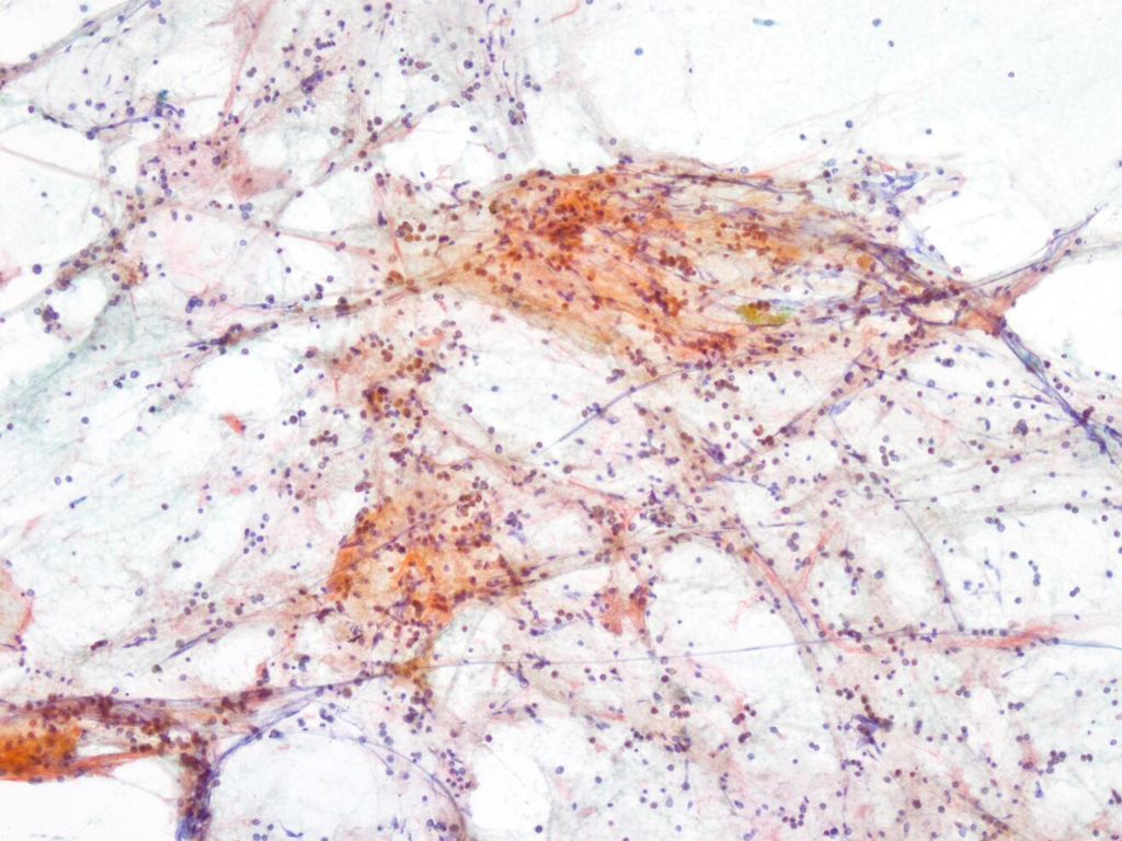
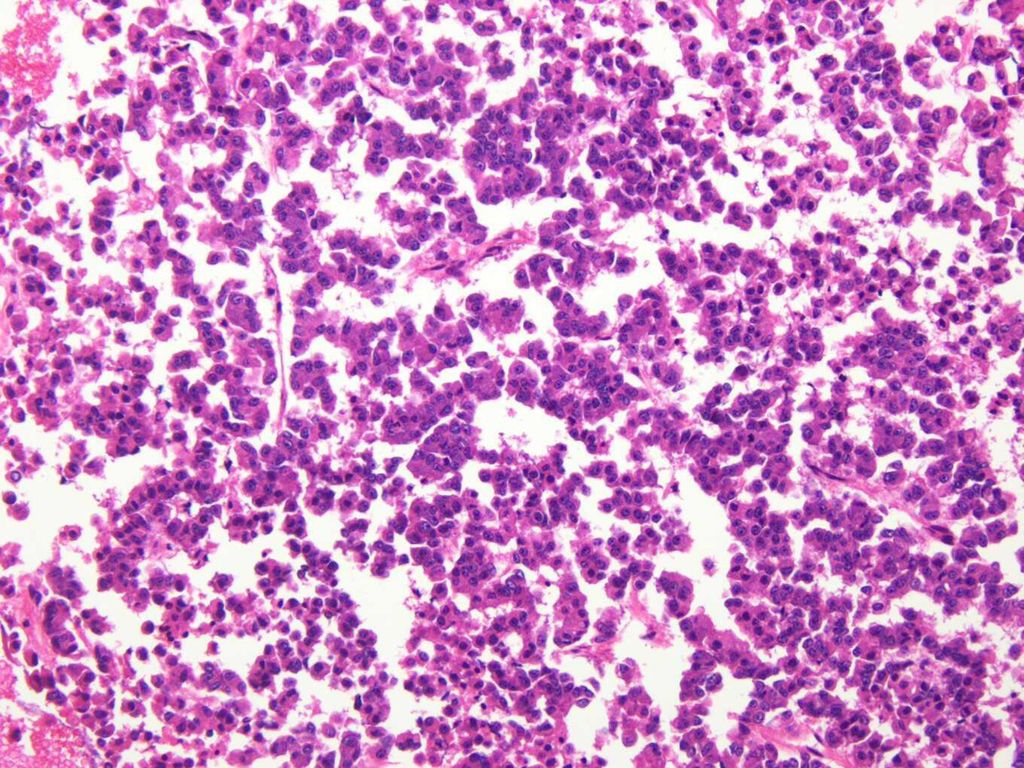
x40

8
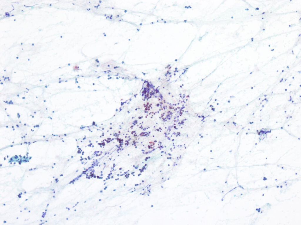
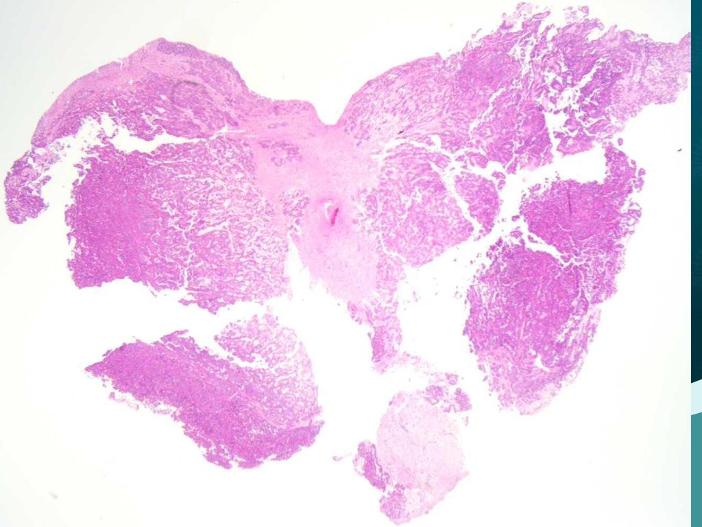
ㅌ100

11
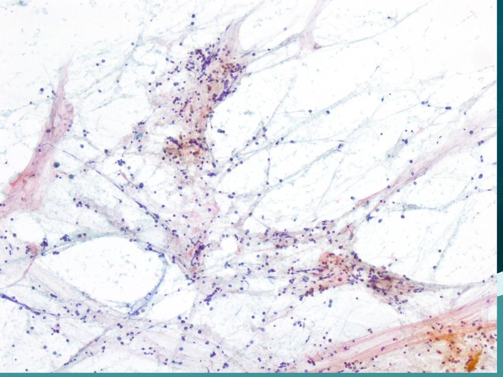
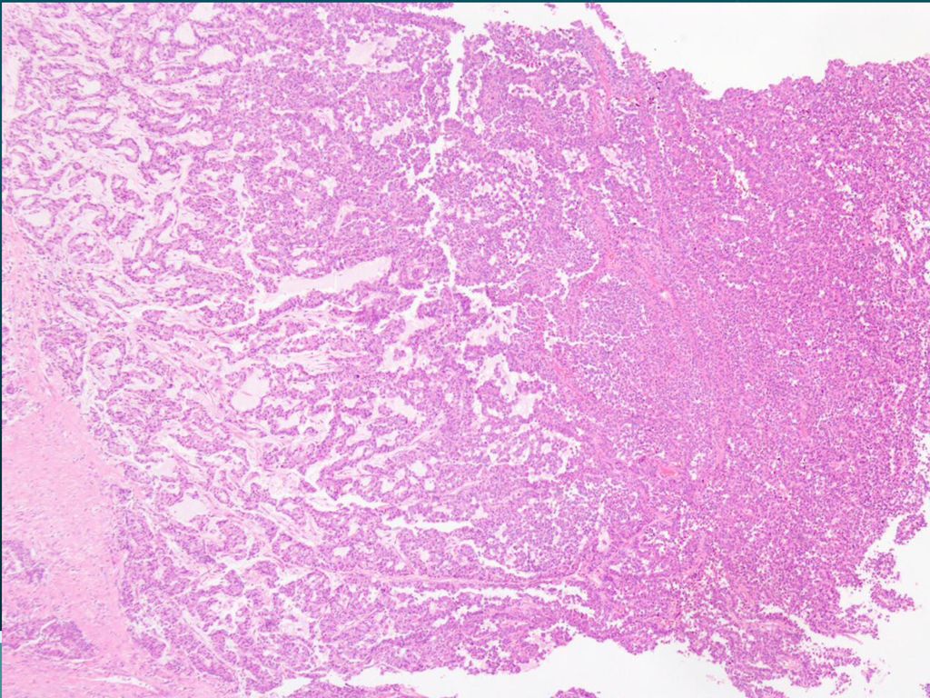
x200

13
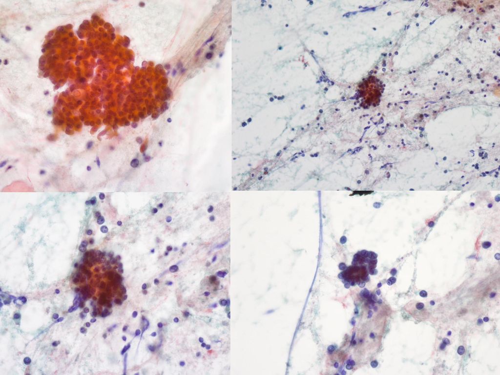
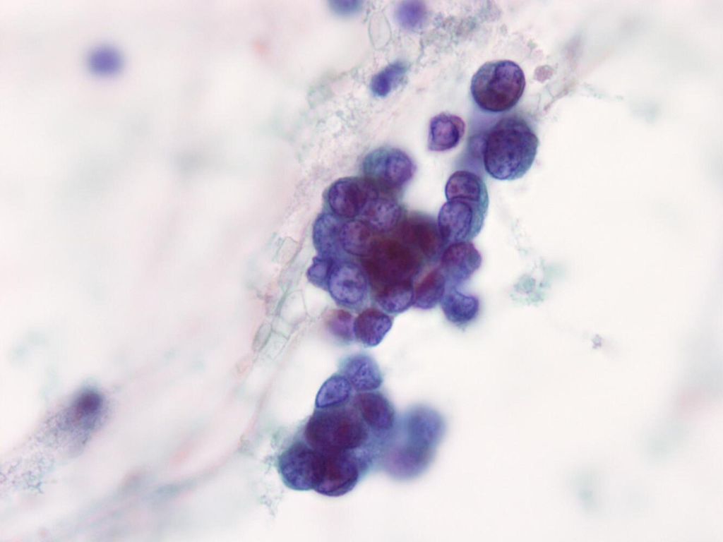
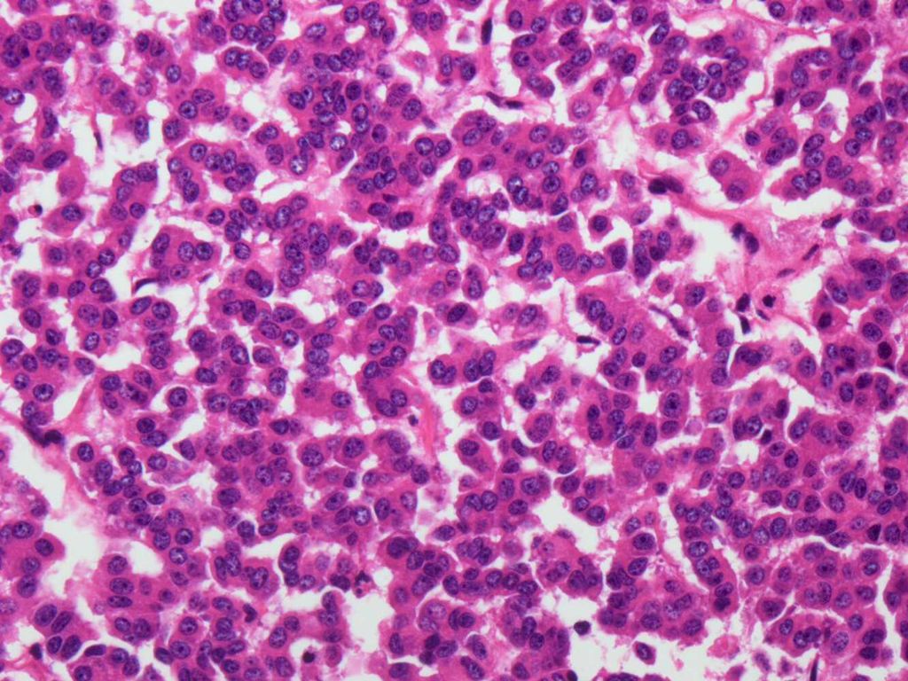
x400

14
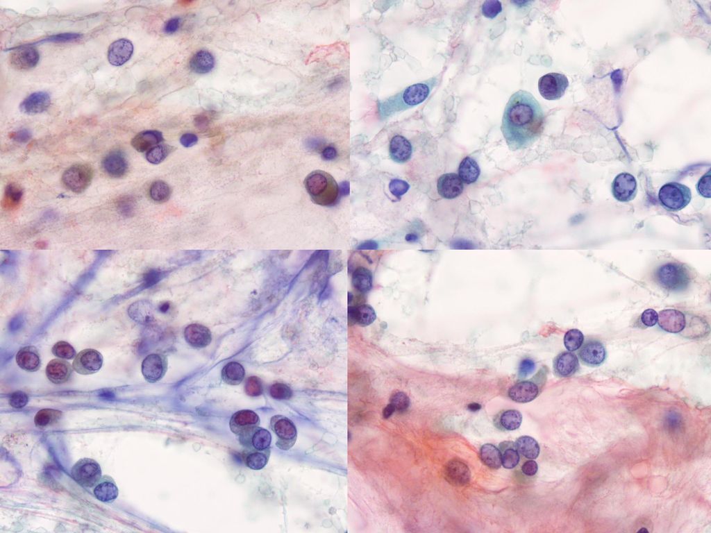
x400

17
Cytologic features No pleomorphism, mitosis, necrosis
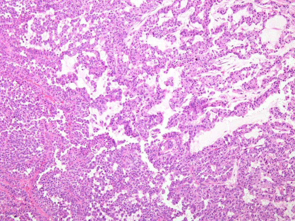
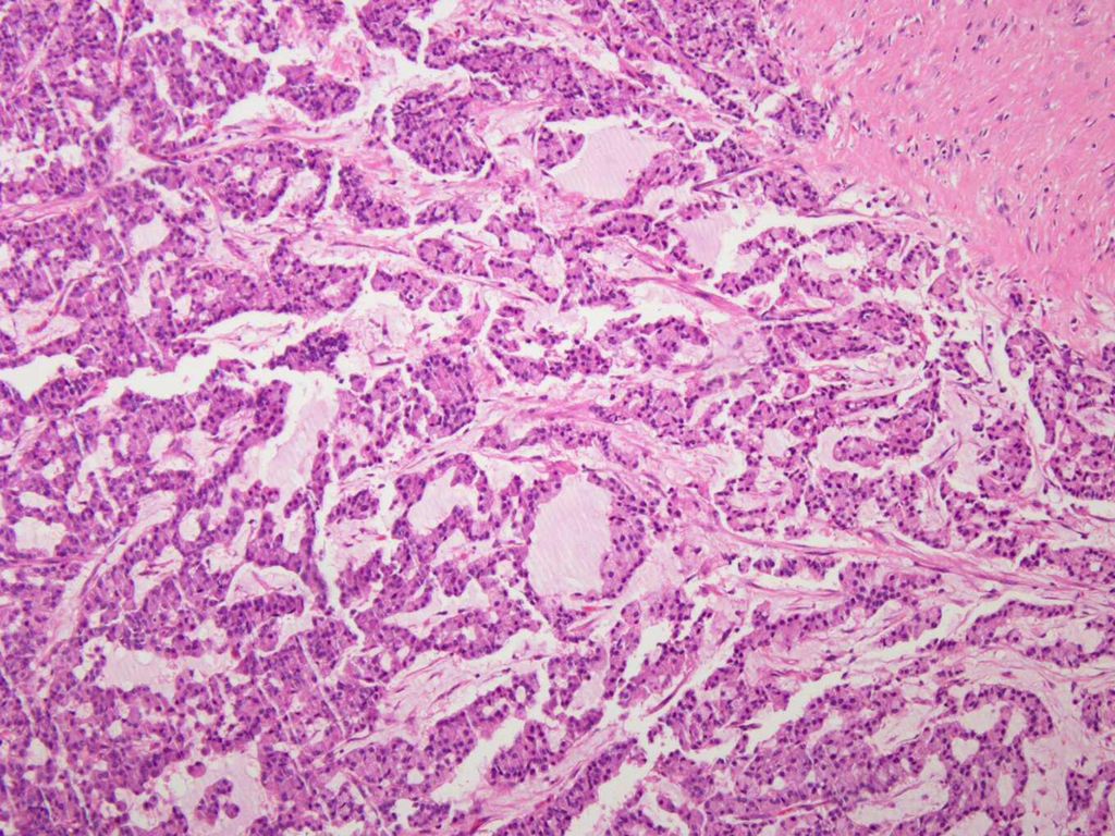
Numerous dispersed individual cells forming infrequently small groups and clusters Fibrovascular connective tissue (delicate branched capillary blood vessel) Tumor cells surrounding, and clinging to, the arborising delicate capillaries Bland, uniform, monotonous small cells with minimal or no variation in cell size Eccentric round nuclei with fine granular chromatin Inconpicuous nucleoli Scanty to moderate basophilic cytoplasm No pleomorphism, mitosis, necrosis
 Tumor cells surrounding, and clinging to, the arborising delicate capillaries. Bland, uniform, monotonous small cells with minimal or no variation in cell size. Eccentric round nuclei with fine granular chromatin. Inconpicuous nucleoli. Scanty to moderate basophilic cytoplasm. No pleomorphism, mitosis, necrosis.")
18
Differential diagnosis
Malignant lymphoma Small cell carcinoma Carcinoid tumor Large cell neuroendocrine carcinoma Adenocarcinoma

19
Differential diagnosis
Malignant lymphoma - no intercellular cohesion - nuclear membrane irregularity - nucleoli+ Small cell carcinoma Carcinoid tumor Large cell neuroendocrine carcinoma Adenocarcinoma

20
Differential diagnosis
Small cell carcinoma - more irregular nuclear contour - pyknosis, necrotic debris - nuclear molding - necrotic background - hyperchromasia - many naked nuclei and nuclear streaking Carcinoid tumor Large cell neuroendocrine carcinoma Adenocarcinoma

21
Differential diagnosis
Large cell neuroendocrine carcinoma - nuclear peomorphism - cohesive cells - fine chromatin - prominent nucleoli - increased mitotic rate - necrotic background Adenocarcinoma Carcinoid tumor

22
Differential diagnosis
Adenocarcinoma -more cytologic atypia -mucin production Carcinoid tumor Small cell carcinoma Malignant lymphoma Large cell neuroendocrine carcinoma

31
Mucicarmine stain

32
Ttf-1 cd56

33
TTF-1 CD56 Ttf-1 cd56

34
TTF-1 CD56 Chromogranin Synaptophysin Ttf-1 cd56

35
Cytologic diagnosis Carcinoid tumor

37
Ki-67

38
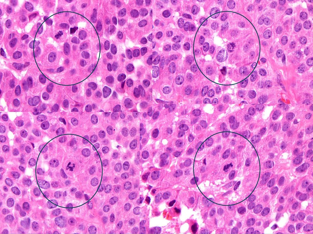
Atypical carcinoid tumor
Final diagnosis Atypical carcinoid tumor

39
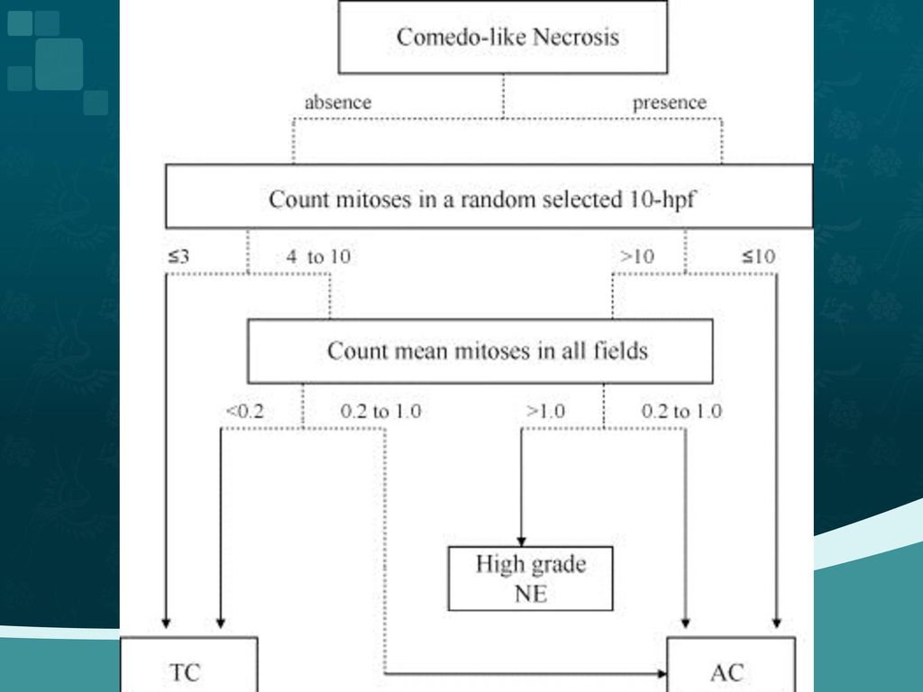
Pathologic criteria of neuroendocrine neoplasm of lung
Typical carcinoid Atypical carcinoid Small-cell lung cancer Large-cell neuroendocrine carcinoma Light microscopic morphology Neuroendocrine morphology (organoid nesting, palisading, rosettes, trabeculae) Neuroendocrine morphology Small size (less than the diameter of 3 small lymphocytes) 1.Scant cytoplasm 2. finely granular chromatin, absent nuclei or faint nucleoli 1.Neuroendocrine morphology 2. Cytologic features of non-small cell carcinoma: large cell size, vesicular, coarse or fine chromatin and/or nucleoli 3.Positive IHC stain for one or more NE markers or neuroendocrine granules by EM, Mitoses per 2mm2 <2 2-10 and/or foci of necrosis >10 Necrosis no Often punctate Frequent, large zone Often large zones Histologic grade Low Intermediated High
 Neuroendocrine morphology. Small size (less than the diameter of 3 small lymphocytes) 1.Scant cytoplasm. 2. finely granular chromatin, absent nuclei or faint nucleoli. 1.Neuroendocrine morphology. 2. Cytologic features of non-small cell carcinoma: large cell size, vesicular, coarse or fine chromatin and/or nucleoli. 3.Positive IHC stain for one or more NE markers or neuroendocrine granules by EM, Mitoses per 2mm2. < and/or foci of necrosis. >10. Necrosis. no. Often punctate. Frequent, large zone. Often large zones. Histologic grade. Low. Intermediated. High.")
42
Cytologic diagnosis and differential diagnosis of lung carcinoid tumors a retrospective study of 63 Cases with histologic correlation. Cancer Cytopathol Dec 25;118(6): 63 cases: cytology with corresponding surgical material cytology specimens : 49 -FNA specimens 14 -brushings/washings. Discordant cases (31 cases,49%) - Overdiagnosis as small cell carcinoma (4 cases; 6%) adenoid cystic tumor (4 cases; 6%) poorly differentiated carcinoma with NE features (5 cases; 8%) - Misdiagnosis of other lesions as TC (4 cases; 6%)
: cases: cytology with corresponding surgical material. cytology specimens : 49 -FNA specimens. 14 -brushings/washings. Discordant cases (31 cases,49%) - Overdiagnosis as small cell carcinoma (4 cases; 6%) adenoid cystic tumor (4 cases; 6%) poorly differentiated carcinoma with NE. features (5 cases; 8%) - Misdiagnosis of other lesions as TC (4 cases; 6%)")
43
Cytologic diagnosis and differential diagnosis of lung carcinoid tumors a retrospective study of 63 Cases with histologic correlation. The significant morphologic factors : nuclear features, chromatin patterns, nucleoli. Nuclear molding and crowding: not discernible features Crush artifact : low and high grade Ki-67 staining: useful

44
Variety of histologic patterns and tumor cells
trabecular, palisading, rosette-like, spindle, glandular, papillary oncocytic, melanin-rich, mucin-producing, signet ring , clear, acinic-like, pleomorphic, bizarre Stromal component: psammomatous, ossification, amyloid deposition

45
경청해주셔서 감사합니다.

Similar presentations
. CC: Low abd distension for 1 M CC: Low abd distension for 1 M (Pap: N these day, CA125: 15) (Pap: N these day, CA125: 15) Wt loss.>")


 제출자 발표 2012.02.01 서울성모병원 병리과 전공의 이영섭.>")
 2) 3) 4) 2. 1) 2) 3. 1) 2) 3) * 혈액의 생성과 파괴 혈구생성부위 -- 1. 2. 3. * 조혈자극인자 –- 1. 2. 3. * 적혈구의 분화와 성숙과정 – Pluripotent stem cell →>")


.>")
>")


 2010. 03. 12. 흉부외과 최 주 원.>")