Download presentation
Presentation is loading. Please wait.

1
Imaging Modalities in Wounds and Superficial Skin Infections

2
28세 여자로 좌측 발바닥 상처에서 고름이 나온다고 내원하였다. 3주 전, 집 마당에서 맨발로 걷다가 나뭇가지를 밟았다고 한다
28세 여자로 좌측 발바닥 상처에서 고름이 나온다고 내원하였다. 3주 전, 집 마당에서 맨발로 걷다가 나뭇가지를 밟았다고 한다. 당시 근처 병원에서 파상풍 주사를 맞았으나 이후로 계속 통증이 발생하여 항생제 처방을 추가로 받았다. 항생제 처방 며칠 후에도 증세는 호전이 안되고 고름이 나오기 시작 했다.

3
내원 당시 이물감은 없었고 주위에 발적이 동반된 2cm 정도의 상처가 있었다. 봉와직염 소견은 없었다. x-ray 촬영 후 항생제 처방하여 외래 f/u 2) I&D 시행 3) 이물질을 찾기 위해 국소 마취 후 개봉 4) 이물질을 찾기 위해 CT를 촬영
 이물질을 찾기 위해 국소 마취 후 개봉. 4) 이물질을 찾기 위해 CT를 촬영.")
4
38% of FB in hand wounds were missed by the initial treating physician
Missed FB are the second leading cause of lawsuits in EM History may be in question Physical examination is also frequently unreliable

5
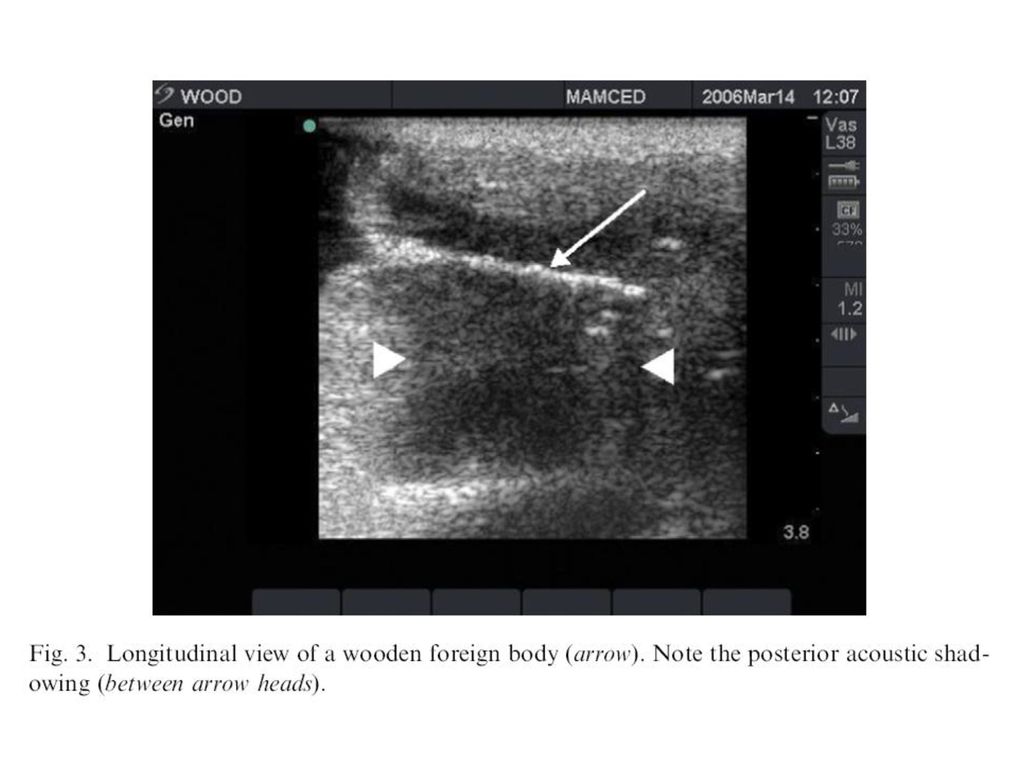
Detection of wood is especially important because it may serve as an unrecognized nidus for infection Cellulitis, abscess, or fistula formation

6
Plain Radiograph Radiopaque substances ≥1-2 mm in size are reliably detected 80-95% of the time Poor sensitivity in detection of radiolucent FB (wood, plastic, vegetative material)
")
7
CT Detect radiopaque and radiolucent FB, with much higher sensitivity for radiopaque ones Given its increased cost, need for ionizing radiation, lack of sensitivity, CT is not routinely recommended to rule out a radiolucent FB

8
CT Gold standard in detection of abscesses
CT has also been used to diagnose and evaluate emergent wound infections (necrotizing fasciitis)
")
9
MRI Accurate in detection of FB
Good case is in which it is difficult to differentiate between cellulitis and necrotizing fasciitis High cost and lack of availability Difficult to distinguish FB from adjacent structures (scar tissue, calcifications, and tendons)
")
10
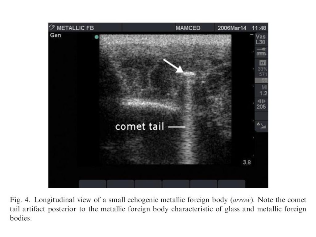
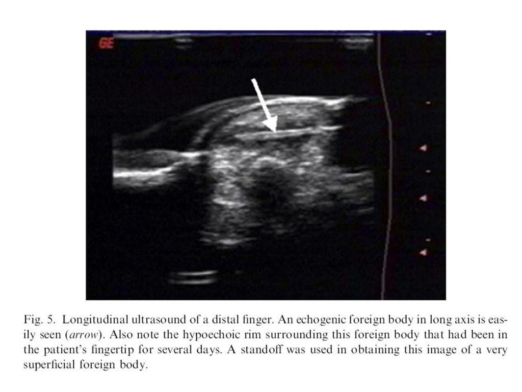
Ultrasound Imaging modality of choice in the evaluation of suspected radiolucent FB

11
High-frequency linear array transducer (≥7.5 MHz)
")
12
Near-field acoustic dead space

13
Standoff pads Commercial products Water-filled latex glove 250- to 500-cc bags of IV fluids

17
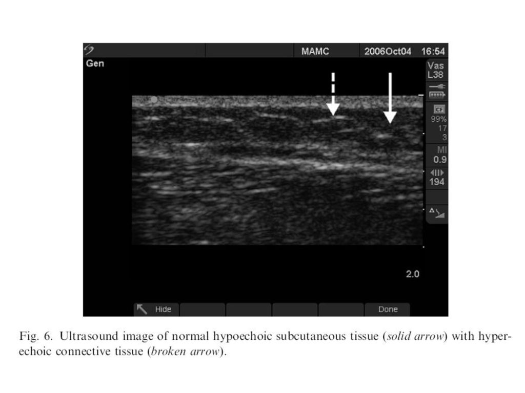
FB are frequently close to normal structures (tendons, muscle, and bone)
Without thorough understanding of anatomy and normal variants a sesamoid bone, scar, or calcification might be mistaken for FB Air in wound is echogenic and may be misinterpreted as FB

18
To carefully examine a wound for FB with US an ample amount of time is required
- Approximately 10 minutes For deeper FB, CT or radiography may be more appropriate

19
US may help the EM provider rapidly differentiate between cellulitis and an abscess
US is 98% sensitive for abscess, whereas PEx is only 86% sensitive

22
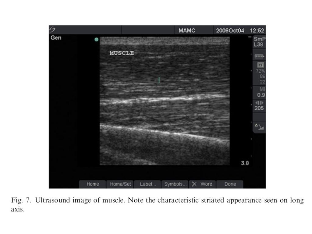
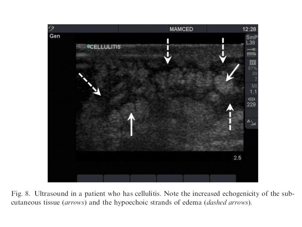
Cellulitis Swelling of SQ tissue Increased distance between the skin and underlying fascia and bone Distant causes diffuse increase in echogenicity of SQ tissue Edema is seen as increased hypoechoic strands between SQ tissue resulting in cobblestone or reticular appearance

24
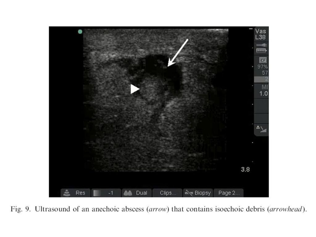
Abscesses have variable appearances
Most common appearance is spherical or elliptical mass Mass is usually hypoechoic or may be anechoic with surrounding echogenic rim Doppler US can be used to look for absence of flow inside the abscess

Similar presentations


![☆ Vocabulary ☆ 단어 및 표현뜻 1. miserable 비참한 2. pimple 여드름 3. treat 치료하다 4. upset 화난 5. huge 거대한, 큰 6. prevent 예방하다 7. soap 비누 8. circular 원형의, 둥근 [Reading]](/40/11054935/big_thumb.jpg "☆ Vocabulary ☆ 단어 및 표현뜻 1. miserable 비참한 2. pimple 여드름 3. treat 치료하다 4. upset 화난 5. huge 거대한, 큰 6. prevent 예방하다 7. soap 비누 8. circular 원형의, 둥근 [Reading]>")


. 가정법에서 if 의 생략 If I should 동사원형 → Should I 동사원형 If I were → Were I If I had pp → Had I pp.>")


