Download presentation
Presentation is loading. Please wait.

1
다양한 신경학적 증상을 보이는 류마티스 환자의 증례
경희대학교 동서 신의학 병원 류마티스 내과 R1 장정윤

2
Case 1 C.C both sole numbness o/s) 2 months ago P.I
박O자 58/F Adm : Case 1 C.C both sole numbness o/s) 2 months ago P.I 3년전에 Rheumatoid Arthritis를 진단 받고 본원 외래 follow up 하는 자로 내원 2개월 전 부터 발생한 상기 증상으로 외래경유 하여 입원 PMHx DM/HTN/Tb/hepatitis (-/-/-/-) FHx. : None PHx. Smoking : None Alcohol : None
 2 months ago. P.I. 3년전에 Rheumatoid Arthritis를 진단 받고 본원 외래 follow up 하는 자로 내원 2개월 전 부터 발생한 상기 증상으로 외래경유 하여 입원. PMHx. DM/HTN/Tb/hepatitis (-/-/-/-) FHx. : None. PHx. Smoking : None. Alcohol : None.")
3
Review of system & Physical exam
Musculoskeletal pain (-) weakness (-) cramp (-) numbness (+) : both sole ,dorsum of foot – 많이 걸으면 따끔 거리는 느낌 동반됨 swelling (-) heating sense (-) Neurologic exam both sole , dorsum sensory : light touch, pin prick; mild decreased motor : dorsiflexion (intact ), plantar flexion (intact ) reflex : ankle jerk, knee jerk ( ++/++ )
 weakness (-) cramp (-) numbness (+) : both sole ,dorsum of foot. – 많이 걸으면 따끔 거리는 느낌 동반됨. swelling (-) heating sense (-) Neurologic exam. both sole , dorsum. sensory : light touch, pin prick; mild decreased. motor : dorsiflexion (intact ), plantar flexion (intact ) reflex : ankle jerk, knee jerk ( ++/++ )")
4
Impression Plan Rheumotoid arthritis combined polyneuropathy
r/o L-spine HIVD Plan Electromyography, Nerve conduction velocity study

5
Initial lab finding CBC/DC /mm² -14.0g/dl – 41.3%- 257K (seg : 69.2 %) Chemistry TB mg/dL AST/ALT /23 U/L ALP/GGT 97/ 23 U/L protein/alb 7.7/4.5 g/dL glucose 100 mg/dL LD /CK / 73 (IU/L) BUN/Cr /0.8mg/dL Uric acid 4.7 mg/dL Na/K/Cl /3.8/104 mmol/L Ca /P / 3.6 mg/dL CRP 0.32 mg/dL ESR 27 mm/h RF > 160 (IU/mL) anti-CCP >100 (U/mL) UA RBC 0-1/HPF WBC 0-1/HPF
 BUN/Cr 17/0.8mg/dL Uric acid 4.7 mg/dL. Na/K/Cl 145/3.8/104 mmol/L Ca /P 10.1 / 3.6 mg/dL. CRP 0.32 mg/dL ESR 27 mm/h. RF > 160 (IU/mL) anti-CCP >100 (U/mL) UA RBC 0-1/HPF WBC 0-1/HPF.")
7
EMG & NCV (2007.4.) 1. Nerve conduction velocity test
- Relativley low conduction in right medial and lateral plantar nerves. - Delayed F-wave latency in right common peroneal nerve. 2. Electromyography normal finding. sensory polyneuropathy bilateral superficial peroneal and right plantar neuropathy 1. 신경전도검사 - Normal motor NCS in bilateral common peroneal and posterior tibial nerves. - Normal sensory NCS in bilateral sural nerves. - - Relativley low SNAPs in right medial and lateral plantar nerves. - Delayed F-wave latency in right common peroneal nerve. - Normal F-waves in other tested nerves. - H-reflexes are demonstrated bilaterally. 2. 침근전도검사 - No denervation potentials in right vastus lateralis, tibialis anterior, peroneus longus, medial head of gastrocnemius, tibialis posterior muscles. - No denervation potentials in bilateral L3-S1 paraspinal muscles. [판정] 전기생리학적 검사상 sensory polyneuropathy (bilateral superficial peroneal and right plantar neuropathy) 를 시사하는 소 견이 관찰됨.
 를 시사하는 소. 견이 관찰됨.")
8
Treatment and Clinical course
Final diagnosis Rheumotoid arthritis combined sensory polyneuropathy Treatment and Clinical course Methylpredinisolone pulse therapy 80% 정도 recovery 되어 외래 follow up 중

9
Case 2 C.C Dysarthria o/s 2개월 전 P.I
박O숙 38/F Adm : Case 2 C.C Dysarthria o/s 2개월 전 P.I 38세 여자 3개월 전 반복되는 구강궤양과 질염으로 Behcet disease를 진단 받고 추적관찰 중 2개월 전부터 시작된 dysarthria, amnesia 증상 있어 외래 경유하여 입원함. PMHx DM/HTN/Tb/hepatitis (-/-/-/-) FHx. : None PHx. Smoking : None Alcohol : None
 FHx. : None. PHx. Smoking : None. Alcohol : None.")
10
Review of system & Physical exam
Head & Neck headache(+) diplopia(-) tinnitus(-) blurred vision (+) : intermittent Neurologic syncope (-) seizure (-) dizziness (-) amensia (+), dysarthria (+) 눈을 감으면 걷기가 불편함 Back & Extremity erythema nodosum in thigh blurred vision : 지난 번 고열 있고 나서 부터 시작 악화 호전 반복하며 심한 두통 동반시 악화되며 당황스러워 지며 더 뿌옇게 보일때가 있다고 함 그러다가도 일 집중하면 잊어버리고 일하고 있는 걸 느낀다고함
11
Impression Plan Behcet disease combined CNS vasculitis
R/O Cerebrovascular accident Behcet uveitis Plan Brain MRI Slit lamp exam to evaluate for uveitis

12
Initial lab finding CBC/DC 7200/mm² -13.5g/dl – 39.6%- 339K (seg :70.6 %) aPTT /34 sec PT(INR) 13.2 (1.01 ) Chemistry TB/DB / mg/dL AST/ALT /12 U/L ALP/GGT 95/ 16 U/L protein/alb 7.6/4.7 g/dL LD /CK / 83 (IU/L) BUN/Cr /0.8mg/dL Uric acid 5.1 mg/dL Na/K/Cl /3.5/101 mmol/L Ca /P / 3.6 mg/dL CRP 0.03 mg/dL ESR 2 mm/h RF 6 (IU/mL)
 BUN/Cr 14/0.8mg/dL Uric acid 5.1 mg/dL. Na/K/Cl 139/3.5/101 mmol/L Ca /P 8.7 / 3.6 mg/dL. CRP 0.03 mg/dL ESR 2 mm/h. RF 6 (IU/mL)")
13
Brain MRI 검사일 : 2007-03-27 판독일 : 2007-03-28 16:38 판독의사 : 배민선/류창우
검사일 : 판독일 : : 판독의사 : 배민선/류창우 검사명 : MRI Br (+) & MRA & Diff [판정] BRAIN MRI and MRA Clinical information: Behcet disease Multifocal areas of T2 signal hyperintensity, bilateral periventricular white matter, internal capsule and basal ganglia . --> possible neuro-Behcet's disease. R/O other vasculitis.
 & MRA & Diff. [판정] BRAIN MRI and MRA. Clinical information: Behcet disease. Multifocal areas of T2 signal hyperintensity, bilateral periventricular white matter, internal capsule and basal ganglia. . --> possible neuro-Behcet s disease. R/O other vasculitis.")
14
Brain MRI 검사일 : 2007-03-27 판독일 : 2007-03-28 16:38 판독의사 : 배민선/류창우
검사일 : 판독일 : : 판독의사 : 배민선/류창우 검사명 : MRI Br (+) & MRA & Diff [판정] BRAIN MRI and MRA Clinical information: Behcet disease Multifocal areas of T2 signal hyperintensity, bilateral periventricular white matter, internal capsule and basal ganglia . --> possible neuro-Behcet's disease. R/O other vasculitis.
 & MRA & Diff. [판정] BRAIN MRI and MRA. Clinical information: Behcet disease. Multifocal areas of T2 signal hyperintensity, bilateral periventricular white matter, internal capsule and basal ganglia. . --> possible neuro-Behcet s disease. R/O other vasculitis.")
15
Treatment & Clinical course
Final diagnosis CNS vasculitis due to Behcet disease Treatment & Clinical course Methylpredinisolone pulse therapy Dysarthria 약간 호전된 상태로 외래 추적 중

16
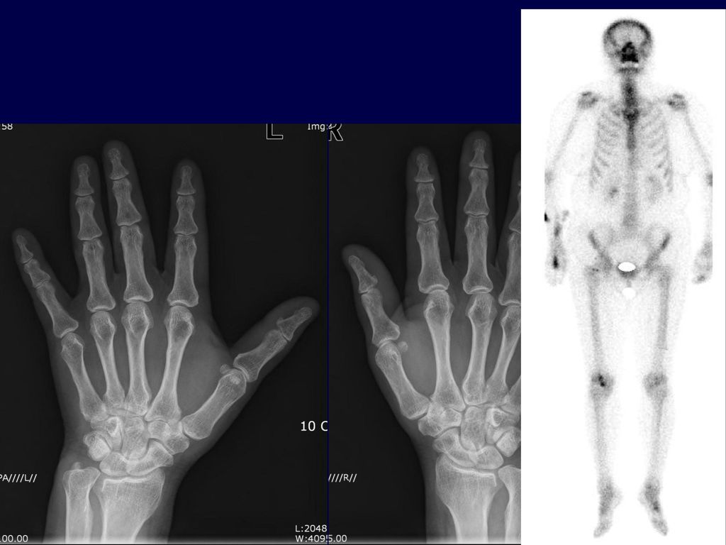
Case 3 C.C Lt 5th finger numbness o/s) 1 month ago P.I
전O남 58/F Adm : Case 3 C.C Lt 5th finger numbness o/s) 1 month ago P.I 58세 여자 환자로 10년전에 혈청음성 류마티스 관절염을 진단 받고 본원 외래 추적 중인 자로 내원 1달 전 부터 시작된 상기 증상으로 외래 경유 하여 입원 Past medical history : none Family history : None Personal history Smoking : None Alcohol : None
 1 month ago. P.I. 58세 여자 환자로 10년전에 혈청음성 류마티스 관절염을 진단 받고 본원 외래 추적 중인 자로 내원 1달 전 부터 시작된 상기 증상으로 외래 경유 하여 입원. Past medical history : none. Family history : None. Personal history. Smoking : None. Alcohol : None.")
17
Review of system & Physical exam
Musculoskeletal & Neurologic arthralgia (+) : both knee, both wrist, both elbow swelling (+) : both ankle numbness (+) : Lt 5th finger tip Back & Extremity CVA tenderness (-/-) pretibial pitting edema (-/-) Tenderness :Rt. shoulder, both elbow, both wrist, Rt. 2nd finger DIP, both ankle
 : both knee, both wrist, both elbow. swelling (+) : both ankle. numbness (+) : Lt 5th finger tip. Back & Extremity. CVA tenderness (-/-) pretibial pitting edema (-/-) Tenderness :Rt. shoulder, both elbow, both wrist, Rt. 2nd finger DIP, both ankle.")
18
Impression Plan Rheumatoid arthritis with R/O peripheral neuropathy
R/O cervical radiculopathy Plan DDx. Spinal cord tumor Medial epicondylitis Recurrent ulnar nerve subluxation Electromyography, Nerve conduction velocity study Prn) MRI
 MRI.")
19
Initial lab finding CBC/DC /mm² -13.4g/dl – 39.7%- 309K (seg : 69.5 %) aPTT /34 sec PT(INR) S54C13.3 (1.02) Chemistry TB/DB / mg/dL AST/ALT /26 U/L ALP/GGT 160/ U/L protein/alb 7.1/4.51g/dL glucose 100 mg/dL LD /CK / 81 (IU/L) BUN/Cr /0.8mg/dL Uric acid 5.7 mg/dL Na/K/Cl /3.6/98 mmol/L Ca /P / 3.4 mg/dL CRP mg/dL ESR 17 mm/h RF 6 IU/mL anti-CCP (-) :0.2 U/mL
 BUN/Cr 18/0.8mg/dL Uric acid 5.7 mg/dL. Na/K/Cl 139/3.6/98 mmol/L Ca /P 9.0 / 3.4 mg/dL. CRP 0.61 mg/dL ESR 17 mm/h. RF 6 IU/mL anti-CCP (-) :0.2 U/mL.")
21
EMG & NCV left ulnar neuropathy on elbow level
(tardy ulnar nerve palsy) [OCR결과/장비결과] 장비인터페이스 [소견] 1. 신경전도검사 - Normal motor NCS in bilateral median and right ulnar nerves. - Low CMAPs above elbow segments in left ulnar nerve by inching technique. - Slow motor NCV across elbow segment in left ulnar nerve. - Relatively low SNAPs in left ulnar nerve, compared with those of right. - Normal sensory NCS in right ulnar, bilateral median and radial superficial sensory nerves. - Normal F-waves in tested nerves. 2. 침근전도검사 - No denervation potentials in left deltoid, triceps brachii, biceps brachii, flexor carpi radialis and ulnaris, and fle xor digitorum profundus (M) muscles. - No denervation potentials in left C5-8, T1 paraspinal muscles. - Mild to moderate denervation potentials in left flexor digitorum profundus (U), abductor digiti minimi, and 1st dorsal interosseous muscles. [판정] 전기생리학적 검사상 left ulnar neuropathy above elbow (tardy ulnar nerve palsy) 를 시사하는 소견이 관찰됨. EMG - General Discussion: - conduction velocity in the ulnar nerve across the elbow is slowed compared with the velocity in the forearm segment; - as noted by Kaempffe et al 1998, abnormal EMG results are associated w/ poor surgical outcome; - position of elbow during EMG/NCS: - remember that elbow flexion may cause greater than a 10% ulnar nerve strain in upto 25% of patients; - it is important to keep elbow flexed 70 to 90 deg during conduction measurements because in this position nerve is optimally stretched; - during extension nerve buckles upon itself & is redundant, factor that may lead to spuriously low computed values of conduction velocity; - amplitude of motor response in abductor digiti minimi is decreased and duration of response is prolonged w/ stimulation above elbow compared w/ stimulation below the elbow; - motor conduction velocity: - look for slowing of more than 10 m/sec; - velocity less than 41 m / sec across the elbos is abnormal; - motor latency from above the elbow more than 10.2 sec; - sensory latency is prolonged; - motor latency: - when axonal degeneration of the ulnar nerve occurs, positive waves and fibrillation may be observed; the proximal edge of the roof of the cubital tunnel was formed by a fibrous band that we call the cubital tunnel retinaculum (CTR). the proximal edge of the roof of the cubital tunnel was formed by a fibrous band that we call the cubital tunnel retinaculum (CTR).
 [OCR결과/장비결과] 장비인터페이스. [소견] 1. 신경전도검사. - Normal motor NCS in bilateral median and right ulnar nerves. - Low CMAPs above elbow segments in left ulnar nerve by inching technique. - Slow motor NCV across elbow segment in left ulnar nerve. - Relatively low SNAPs in left ulnar nerve, compared with those of right. - Normal sensory NCS in right ulnar, bilateral median and radial superficial sensory nerves. - Normal F-waves in tested nerves. 2. 침근전도검사. - No denervation potentials in left deltoid, triceps brachii, biceps brachii, flexor carpi radialis and ulnaris, and fle. xor digitorum profundus (M) muscles. - No denervation potentials in left C5-8, T1 paraspinal muscles. - Mild to moderate denervation potentials in left flexor digitorum profundus (U), abductor digiti minimi, and 1st dorsal. interosseous muscles. [판정] 전기생리학적 검사상 left ulnar neuropathy above elbow (tardy ulnar nerve palsy) 를 시사하는 소견이 관찰됨. EMG - General Discussion: - conduction velocity in the ulnar nerve across the elbow is slowed compared with the velocity in the forearm segment; - as noted by Kaempffe et al 1998, abnormal EMG results are associated w/ poor surgical outcome; - position of elbow during EMG/NCS: - remember that elbow flexion may cause greater than a 10% ulnar nerve strain in upto 25% of patients; - it is important to keep elbow flexed 70 to 90 deg during conduction measurements because in this position nerve is optimally stretched; - during extension nerve buckles upon itself & is redundant, factor that may lead to spuriously low computed values of conduction velocity; - amplitude of motor response in abductor digiti minimi is decreased and duration of response is prolonged w/ stimulation above elbow compared w/ stimulation below the elbow; - motor conduction velocity: - look for slowing of more than 10 m/sec; - velocity less than 41 m / sec across the elbos is abnormal; - motor latency from above the elbow more than 10.2 sec; - sensory latency is prolonged; - motor latency: - when axonal degeneration of the ulnar nerve occurs, positive waves and fibrillation may be observed; the proximal edge of the roof of the cubital tunnel was formed by a fibrous band that we call the cubital tunnel retinaculum (CTR). the proximal edge of the roof of the cubital tunnel was formed by a fibrous band that we call the cubital tunnel retinaculum (CTR).")
22
Elbow MRI , Lt (2007.4.11) [소견] LEFT HUMERUS MRI WITH ENHANCEMENT
1. No gross evidence of mass or cystic lesion along the ulnar nerve pathway or posteromedial aspect of elbow. 2. Joint effusion and synovial enhancement in elbow joint. - with marginal bony spur, prominent esp. in medial portion of elbow joint. --> Degenerative change > RA change. 3. Abrupt and focal thinning of ulnar nerve at oleccaron-humeral joint level, posteromedial aspect. - with scanty amount of surrounding fat or loss of surrounding fat around ulnar nerve. - at level of medial collateral ligament. - with mild degree of surrounding soft tissue enhancement. --> Suggested ulnar neuropathy at elbow level (cubital tunnel syndrome). ; probably due to tightness of retinaculum.
![Elbow MRI , Lt ( ) [소견] LEFT HUMERUS MRI WITH ENHANCEMENT](http://slidesplayer.org/slide/13077805/79/images/22/Elbow+MRI+%2C+Lt+%28+%29+%5B%EC%86%8C%EA%B2%AC%5D+LEFT+HUMERUS+MRI+WITH+ENHANCEMENT.jpg "1. No gross evidence of mass or cystic lesion along the ulnar nerve pathway or posteromedial aspect of elbow. 2. Joint effusion and synovial enhancement in elbow joint. - with marginal bony spur, prominent esp. in medial portion of elbow joint. --> Degenerative change > RA change. 3. Abrupt and focal thinning of ulnar nerve at oleccaron-humeral joint level, posteromedial aspect. - with scanty amount of surrounding fat or loss of surrounding fat around ulnar nerve. - at level of medial collateral ligament. - with mild degree of surrounding soft tissue enhancement. --> Suggested ulnar neuropathy at elbow level (cubital tunnel syndrome). ; probably due to tightness of retinaculum.")
23
Treatment & Clinical course
Final diagnosis Cubital tunnel syndrome Treatment & Clinical course OP 계획 하였으나 환자 refuse 하여 경과 관찰 중

24
Case 4 C.C Hoarseness , swallowing difficulty o/s 1month ago P.I
이O기 50/M Adm : Case 4 C.C Hoarseness , swallowing difficulty o/s 1month ago P.I 한달 전 부터 시작된 상기 증상으로 인근 병원에서 Laryngoscopy, Neck CT 시행한 결과 Lt vocal cord palsy, central type 소견 보여 정밀 검사 위해 본원 신경과 입원함 Past medical history - DM/HTN/Tb/hepatitis (-/-/-/-) PHx. - Smoking(-) - Alcohol (-)
 PHx. - Smoking(-) - Alcohol (-)")
25
Review of system & Physical exam
Head & Neck headache(+) diplopia(-) tinnitus(-), blurred vision (-), hoarseness(+), swallowing difficulty (+) tongue deviation(-) Musculoskeletal Arthralgia: both ankle, both wrist Neurologic exam Mental & Speech: normal Facial palsy (-/-) blurred vision : 지난 번 고열 있고 나서 부터 시작 악화 호전 반복하며 심한 두통 동반시 악화되며 당황스러워 지며 더 뿌옇게 보일때가 있다고 함 그러다가도 일 집중하면 잊어버리고 일하고 있는 걸 느낀다고함
 diplopia(-) tinnitus(-), blurred vision (-), hoarseness(+), swallowing difficulty (+) tongue deviation(-) Musculoskeletal. Arthralgia: both ankle, both wrist. Neurologic exam. Mental & Speech: normal. Facial palsy (-/-) blurred vision : 지난 번 고열 있고 나서 부터 시작. 악화 호전 반복하며. 심한 두통 동반시 악화되며. 당황스러워 지며 더 뿌옇게 보일때가 있다고 함. 그러다가도 일 집중하면 잊어버리고 일하고 있는 걸 느낀다고함.")
26
Impression Plan Brain MRI prn) CSF exam Known vocal cord palsy
R/O due to Cerebral infarction R/O due to CNS infection Plan Brain MRI prn) CSF exam DDx. Spinal cord tumor Medial epicondylitis Recurrent ulnar nerve subluxation
 CSF exam. DDx. Spinal cord tumor. Medial epicondylitis. Recurrent ulnar nerve subluxation.")
27
Initial lab finding CBC/DC /mm g/dl – 35%- 666K/uL (seg :74.4 %) aPTT 35/34 sec PT(INR) 1.15 (1.01 ) Chemistry TB/DB /0.2 mg/dL AST/ALT /35 U/L ALP/GGT 90/ 38 U/L protein/alb 7.4/3.6 g/dL Uric acid: 2.3mg/dL BUN/Cr /0.7mg/dL Na/K/Cl /4.3/97 mmol/L Ca /P 8.5 / 2.9 mg/dL ESR: 93mm/h UA Blood: protein: Microscopic: RBC 5-9/HPF, WBC: 5~9/HPF Autoantibody ANA speckled(1+) 1:160 Anti-ds-DNA IgG : Positive (57.2 IU/mL) ‥(응급)Neutrophil segment% 74.4 (%) ▲ 40 ~ 60 ‥(응급)Lymphocyte% 13.0 (%) ▼ 20 ~ 50 ‥(응급)Monocyte% 6.6 (%) 2 ~ 10 ‥(응급)Eosinophil% 5.0 (%) ▲ 0 ~ 4 ‥(응급)Basophil% 1.0 (%) 0 ~ 1
 1:160 Anti-ds-DNA IgG : Positive (57.2 IU/mL) ‥(응급)Neutrophil segment% 74.4 (%) ▲ 40 ~ 60. ‥(응급)Lymphocyte% 13.0 (%) ▼ 20 ~ 50. ‥(응급)Monocyte% 6.6 (%) 2 ~ 10. ‥(응급)Eosinophil% 5.0 (%) ▲ 0 ~ 4. ‥(응급)Basophil% 1.0 (%) 0 ~ 1.")
28
Brainstem auditory evoked potential
Brain MRI 특이 소견 관찰 되지 않음. Brainstem auditory evoked potential Upper brainstem lesion between lower pons and midbrain 을 시사하는 소견이 관찰됨.

29
‥(응급)Neutrophil segment% 74.4 (%) ▲ 40 ~ 60
‥(응급)Lymphocyte% 13.0 (%) ▼ 20 ~ 50 ‥(응급)Monocyte% 6.6 (%) 2 ~ 10 ‥(응급)Eosinophil% 5.0 (%) ▲ 0 ~ 4 ‥(응급)Basophil% 1.0 (%) 0 ~ 1
Lymphocyte% 13.0 (%) ▼ 20 ~ 50. ‥(응급)Monocyte% 6.6 (%) 2 ~ 10. ‥(응급)Eosinophil% 5.0 (%) ▲ 0 ~ 4. ‥(응급)Basophil% 1.0 (%) 0 ~ 1.")
30
(American Rheumatism association)
Diagnostic criteria (American Rheumatism association) 1) Malar rash 2) Discoid rash 3) photosensitivity 4) Oral ulcers 5) Arthritis: nonerosive arthritis involving 2 or more peripheral joint 6) Serositis : pleuritis, pericarditis 7) Renal disorder: persistent proteinuria, cellular casts 8) Neurologic disorder : seizures or psychosis 9) Hematologic disorder : hemolytic anemia/ leukopenia / lymphopenia / thrombocytopenia 10) Immunologic disorders: antiphophlipid Ab or anti-dsDNA Ab or anti-Sm or VDRL 11) ANA
 1) Malar rash. 2) Discoid rash. 3) photosensitivity. 4) Oral ulcers. 5) Arthritis: nonerosive arthritis involving 2 or more peripheral joint. 6) Serositis : pleuritis, pericarditis. 7) Renal disorder: persistent proteinuria, cellular casts. 8) Neurologic disorder : seizures or psychosis. 9) Hematologic disorder : hemolytic anemia/ leukopenia / lymphopenia / thrombocytopenia. 10) Immunologic disorders: antiphophlipid Ab or anti-dsDNA Ab or anti-Sm or VDRL. 11) ANA.")
31
Final Diagnosis Plan Probable SLE with CNS neuropathy
MPD pulse 1g .day 5 days Methylpredinisolone pulse therapy L-tube feeding 하면서 외래 추적 관찰 중

Similar presentations


 3 days ago CC: Feveronset) 3 days ago PI : F/29, 5 년전 첫아기 임신 8 개월째에.>")
 인체기능 – 생리학 (Physiology) 생리학 인체의 세포, 조직 또는 각 기관이나 계통 등과 같은 인체구성단위 들이 생명현상을.>")


신경발달치료(NDT)>")


 근 골격계 사정 (1) 관절 가동범위 측정 시진 : 양쪽 대칭, 상 하지 비교, 위축 관절과 뼈의 변형, 근육 모양, 크기 촉진 : 경련, 근육의 긴장도, 압통 관절 각도계 : 객관적.>")