Download presentation
Presentation is loading. Please wait.

1
유전자의 구조 Structures of Genes
생물환경학과 김 정 호 Department of Bio-Environmental Science

2
유전 (Heredity) 멘델의 유전법칙 (Mendel’s Genetic Laws)
Gregor Mendel (1822–1884) Austrian monk 1856 ~ 1863 : 29,000 pea plants Father of Genetics Mendelian Inheritance 분리의 법칙 (Law of Segregation) 독립의 법칙 (Law of Independent Assortment) 우열의 원리 (Principle of Dominance) 멘델 법칙의 재발견 (Rediscovery of Mendel’s Genetic Laws) . 1900 . Hugo de Vries, Carl Correns, Erich von Tschermak
 Austrian monk ~ 1863 : 29,000 pea plants. Father of Genetics. Mendelian Inheritance. 분리의 법칙 (Law of Segregation) 독립의 법칙 (Law of Independent Assortment) 우열의 원리 (Principle of Dominance) 멘델 법칙의 재발견 (Rediscovery of Mendel’s Genetic Laws) Hugo de Vries, Carl Correns, Erich von Tschermak.")
3
유전자와 염색체 (Gene & Chromosome)
Wilhelm Johannsen (Danish botanist) the fundamental physical and functional units of heredity 염색체설 (Chromosomal Theory of Heredity) 1910, Thomas Hunt Morgan (1866 ~ 1945) Nobel Prize in Physiology or Medicine, 1933 염색체가 유전의 본질 Genes reside on specific chromosomes Genes occupy specific locations on the chromosome the first chromosomal map of the fruit fly Drosophila melanogaster .
 the fundamental physical and functional units of. heredity. 염색체설 (Chromosomal Theory of Heredity) 1910, Thomas Hunt Morgan (1866 ~ 1945) Nobel Prize in Physiology or Medicine, 염색체가 유전의 본질. Genes reside on specific chromosomes. Genes occupy specific locations on the chromosome. the first chromosomal map of the fruit fly. Drosophila melanogaster. .")
4
유전물질 (Genetic Material)
Protein vs DNA Proten : 20 amino acids DNA : 4 bases (A, T, G, C)
")
5
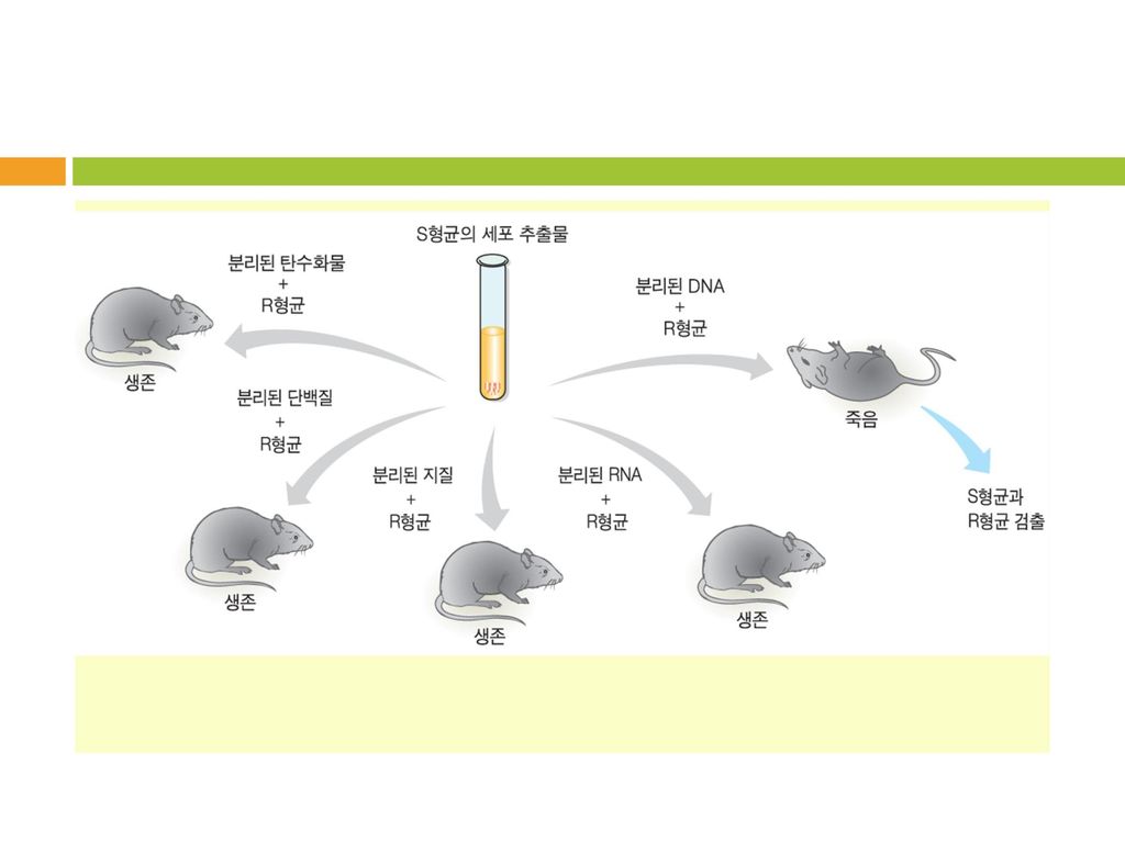
Griffith의 형질전환 실험 1928, Frederick Griffith (1877–1941, UK)
폐렴 쌍구균 (Streptococcus pneumoniae) S (smooth) : with capsule, 병원성 (pathogenic) R (rough) : without capsule, 비병원성 (non-pathogenic) 비병원성 (R) → 병원성 (S) : 형질전환 (Transformation) the first demonstrations of bacterial transformation (in vivo) 형질전환의 본질 (Transforming principle) ?
 S (smooth) : with capsule, 병원성 (pathogenic) R (rough) : without capsule, 비병원성 (non-pathogenic) 비병원성 (R) → 병원성 (S) : 형질전환 (Transformation) the first demonstrations of bacterial transformation. (in vivo) 형질전환의 본질 (Transforming principle)")
6
Griffith's experiment

7
Avery–MacLeod–McCarty Experiment
1944, Oswald Avery, Colin MacLeod, Maclyn McCarty DNA is the transforming principle (hereditary material)
")
9
Hershey–Chase experiments
1952, Alfred Hershey and Martha Chase Showed that when bacteriophages, which are composed of DNA and protein, infect bacteria, their DNA enters the host bacterial cell, but most of their protein does not. Helped to confirm that DNA is the genetic material. Hershey shared the 1969 Nobel Prize in Physiology or Medicine with Max Delbrück and Salvador Luria for their “discoveries concerning the genetic structure of viruses.

10
Hershey–Chase : Blender experiments, 1952

11
핵산의 발견 1896, Johannes Friedrich Miescher (1844~1895)
Swiss physician & biologist from the nuclei of white blood cells (pus) nuclein : 세포의 핵(nucleus) 안에서 발견 phosphate-rich
 nuclein : 세포의 핵(nucleus) 안에서 발견. phosphate-rich.")
12
핵산의 구성성분 Nucleic acid DNA : deoxyribonucleic acid
RNA : ribonucleic acid 기본 단위 : nucleotide 당(sugar), 염기(base), 인산(phosphate) 당 5탄당 (pentose) RNA : ribose DNA : deoxyribose
, 염기(base), 인산(phosphate) 당. 5탄당 (pentose) RNA : ribose. DNA : deoxyribose.")
13
염기 (Bases)
")
14
Nucleoside, Nucleotide

15
Nucleoside (Deoxynucleoside) : 당 + 염기
RNA : adenosine, guanosine, cytidine, uridine DNA : deoxyadenosine, deoxyguanosine, deoxycytidine, deoxythymidine Nucleotide(Deoxynucleotide) : nucleoside + 인산 Nucleoside monophosphate RNA : AMP(adenosine monophosphate) UMP, GMP, CMP DNA : dAMP(deoxyadnosine monophosphate), dTMP, dGMP, dCMP Nucleoside diphosphate : ADP (adenosine diphosphate) Nucleoside triphosphate : ATP (adenosine triphosphate)
 : nucleoside + 인산. Nucleoside monophosphate. RNA : AMP(adenosine monophosphate) UMP, GMP, CMP. DNA : dAMP(deoxyadnosine monophosphate), dTMP, dGMP, dCMP. Nucleoside diphosphate : ADP (adenosine diphosphate) Nucleoside triphosphate : ATP (adenosine triphosphate)")
16
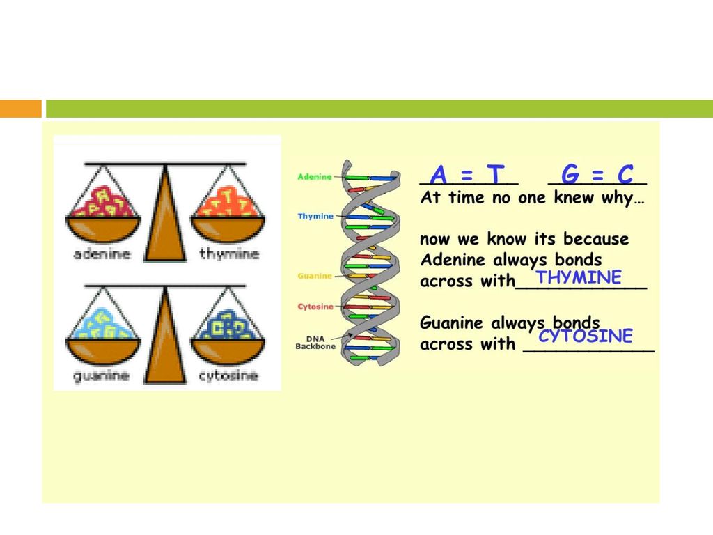
Chargaff’s Rule 1950s, Erwin Chargaff (1905 ~ 2002)
Austro-Hungarian biochemist Columbia University (Medical School)
")
18
1st rule : Base Pair Rule DNA from any cell of all organisms should have a 1:1 ratio of pyrimidine and purine bases. More specifically, the number of guanine units equals the number of cytosine units, and the number of adenine units equals the number of thymine units. This hinted at the base pair makeup of DNA. 2nd rule The relative amounts of G, C, A, and T bases varies from one species to another. This hinted that DNA rather than protein could be the genetic material.

19
X-ray Diffraction Analysis : photo 51
1952 Raymond Gosling, Rosalind Franklin, Maurice Wilkins

20
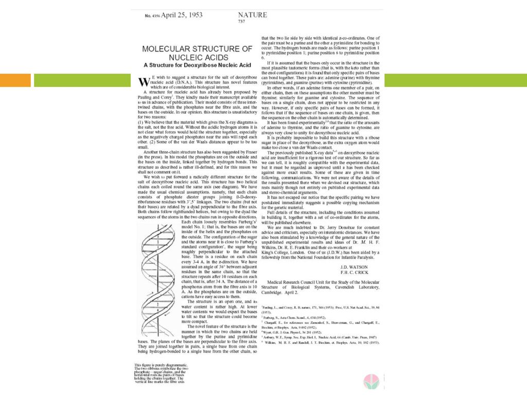
James Wastson & Francis Crick
DNA Double Helix 1953(23, 34) James Wastson & Francis Crick
 James Wastson & Francis Crick.")
22
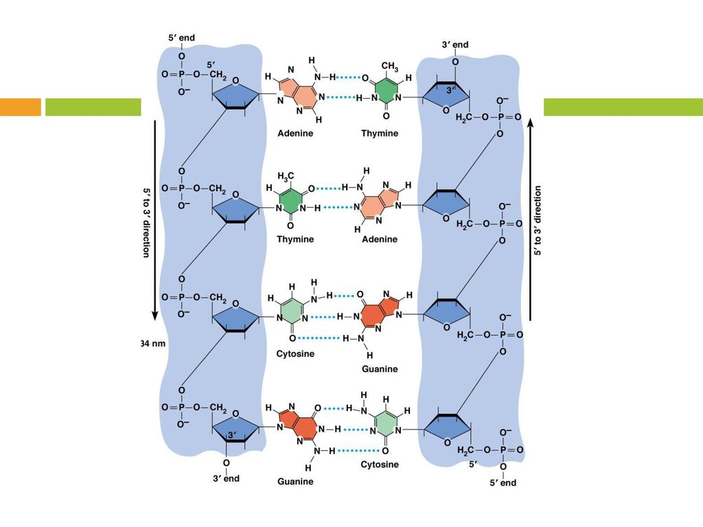
Polynucleotide 당-인산 골격 (sugar-phosphate backbone) 인산다이에스터 결합
Nucleotide와 nucleotide가 phosphodiester bond 방향성 : 5’ → 3’ 5’ end : phosphate group 3’ end : hydroxyl group

23
DNA Double Helix (B-DNA)
")
25
Double-stranded helix
나선 바깥쪽 : Sugar-Phosphate Backbone 나선 내부 : 염기 쌍 (purine–pyrimidine) Complementary base pairing (상보적인 염기 쌍 형성) A-T : 2 hydrogen bonds G-C : 3 hydrogen bonds Antiparallel (역평행) 5’ >3’ 3’< ’
 Complementary base pairing (상보적인 염기 쌍 형성) A-T : 2 hydrogen bonds. G-C : 3 hydrogen bonds. Antiparallel (역평행) 5’ >3’ 3’< ’")
26
A-, B-, Z-form DNA A-form B-form Z- form

27
B-form DNA Watson-Crick Right-handed double helix The most common form, at neutral pH & physiological salt concentrations Z-form DNA Left-handed double helix Zig-Zag pattern in the phosphodiester backbone A small amount of the DNA in a cell A-form nucleic acid Right-handed duplex for RNA-DNA duplexes & RNA-RNA duplexes

28
DNA 복제 (DNA Replication)
복제개시점(origin of replication, ori ) 반보존적 복제(semiconservative replication) DNA 중합효소(DNA polymerase) primer dNTP(dATP, dTTP, dGTP, dCTP) 합성은 5’ → 3’ 방향으로 진행 양방향(bidirectional replication) 반불연속적(semidiscontinuous) High fidelity : 교정 기능 (proofreading)
 반보존적 복제(semiconservative replication) DNA 중합효소(DNA polymerase) primer. dNTP(dATP, dTTP, dGTP, dCTP) 합성은 5’ → 3’ 방향으로 진행. 양방향(bidirectional replication) 반불연속적(semidiscontinuous) High fidelity : 교정 기능 (proofreading)")
29
반보존적 복제 (Semiconservative Replication)
")
30
Meselson-Stahl Experiment
1958, Matthew Meselson and Franklin Stahl

31
복제의 시작 DNA topoisomerase unwinding of DNA superhelix DNA helicase
binding to the origin of replication ori (복제 개시점, 복제 원점) unwinding and separation of the two strands of the double helix Single strand binding protein SSBP (단일가닥 결합 단백질) stabilization of separated strands
 unwinding and separation of the two strands. of the double helix. Single strand binding protein. SSBP (단일가닥 결합 단백질) stabilization of separated strands.")
32
DNA 중합효소 (DNA polymerase)
DNA polymerase (DNA-dependent DNA polymerase) 주형 가닥에 상보적인 딸 가닥 합성 → 반보존적 복제 (semiconservative replication) 시발체 (primer)가 필요함 합성은 5’→ 3’방향으로 효소가 주형 가닥을 3’ 방향에서 5’방향으로 읽어 나감 선행 nucleotide인 DNA 가닥의 3’-OH 말단에 더해지는 nucleotide의 5’-PO4-3 가 결합함 phosphodiester bond 형성 에너지가 필요함 : dNTP (dATP, dTTP, dGTP, dCTP)
 주형 가닥에 상보적인 딸 가닥 합성. → 반보존적 복제 (semiconservative replication) 시발체 (primer)가 필요함. 합성은 5’→ 3’방향으로. 효소가 주형 가닥을 3’ 방향에서 5’방향으로 읽어 나감. 선행 nucleotide인 DNA 가닥의 3’-OH 말단에. 더해지는 nucleotide의 5’-PO4-3 가 결합함. phosphodiester bond 형성. 에너지가 필요함 : dNTP (dATP, dTTP, dGTP, dCTP)")
33
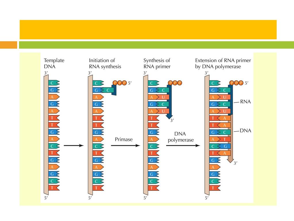
시발체 (Primer) 시발체 (Primer) Short RNA segment complementary to ss DNA
template DNA primase : a type of RNA polymerase (DNA – RNA) 복제 후 DNA polymerase I (5’-to-3’ exonuclease)에 의해 제거됨
 복제 후 DNA polymerase I (5’-to-3’ exonuclease)에. 의해 제거됨.")
35
복제는 5’→ 3’방향으로

36
양방향 복제 (Bidirectional Replication)
")
37
Leading strand vs Lagging strand
DNA : Antiparallel (역평행) 5’ >3’ 3’< ’ DNA 합성 : 5’ → 3’ Leading strand (선도가닥) : 연속적 합성 중합효소가 복제분기점(Replication fork) 쪽으로 진행 Lagging strand (지연가닥) : 불연속적 합성 중합효소가 복제분기점 반대 방향으로 진행 Okazaki fragment DNA polymerase I : RNA primer 제거 DNA ligase(연결효소) : Okazaki fragment 연결
 5’ >3’ 3’< ’ DNA 합성 : 5’ → 3’ Leading strand (선도가닥) : 연속적 합성. 중합효소가 복제분기점(Replication fork) 쪽으로 진행. Lagging strand (지연가닥) : 불연속적 합성. 중합효소가 복제분기점 반대 방향으로 진행. Okazaki fragment. DNA polymerase I : RNA primer 제거. DNA ligase(연결효소) : Okazaki fragment 연결.")
38
반불연속적 복제 (Semi-discontinuous replication)
")
39
원핵세포, 진핵세포의 DNA 복제

40
1 유전자 1 효소설 One gene-One Enzyme hypothesis
George Beadle & Edward Tatum, 1941 Neurospora crassa (붉은 빵곰팡이) Auxotrophic mutants (영양요구 돌연변이주) X-ray, arginine 요구 1958 Nobel prize Genes act through the production of enzymes. Each gene is responsible for producing a single enzyme. Each enzyme affects a single step in a metabolic pathway. One gene-One Polypeptide hypothesis
 Auxotrophic mutants (영양요구 돌연변이주) X-ray, arginine 요구 Nobel prize. Genes act through the production of enzymes. Each gene is responsible for producing a single enzyme. Each enzyme affects a single step in a metabolic. pathway. One gene-One Polypeptide hypothesis.")
41
유전정보(생명)의 중심원리 Central Dogma of Life (Molecular Biology)
Francis Crick, 1958 (1970) 생명체 내에서 유전정보의 흐름 Flow of genetic information within a biological system 전사 (transcription, DNA → RNA) : RNA polymerase 번역 (translation, RNA → Protein) : Ribosome, tRNA 복제 (replication (DNA → DNA) : DNA polymerase Exceptions : 역전사 (reverse transcription, RNA → DNA) : Reverse transcriptase RNA 복제 (RNA replication,RNA → RNA) ; RNA-dependent RNA polymerase
 생명체 내에서 유전정보의 흐름. Flow of genetic information within a biological system. 전사 (transcription, DNA → RNA) : RNA polymerase. 번역 (translation, RNA → Protein) : Ribosome, tRNA. 복제 (replication (DNA → DNA) : DNA polymerase. Exceptions : 역전사 (reverse transcription, RNA → DNA) : Reverse transcriptase. RNA 복제 (RNA replication,RNA → RNA) ; RNA-dependent RNA polymerase.")
42
유전자 (Gene) 유전자 (Gene) DNA의 한 부분으로 유전의 단위 물리적으로 구별되어 있지는 않음
구조 유전자 (structural gene) RNA 또는 Protein 암호화 조절 유전자 (control gene) 발현 조절
 RNA 또는 Protein 암호화. 조절 유전자 (control gene) 발현 조절.")
43
원핵/진핵 세포의 유전자 구조 원핵세포 (Procaryotic cell) No exon/intron
Polycistronic : Operon 구성 하나의 promoter에 의해 여러 개의 구조유전자 조절 cistron : 구조 유전자에서 한 polypeptide의 아미노산 서열을 결정하는 유전자 단위 진핵세포 (Eucaryotic cell) Exon / Intron : intron은 RNA splicing에 의해 제거 (mature mRNA) Monocistronic : 하나의 promoter에 하나의 구조유전자 연결
 Exon / Intron : intron은 RNA splicing에 의해 제거 (mature mRNA) Monocistronic : 하나의 promoter에 하나의 구조유전자 연결.")
44
원핵세포의 유전자 구조

45
진핵세포의 유전자 구조

Similar presentations


.>")


이란? 게놈(genome)은 유전자(gene)와 염색체(chromosome)>")