Download presentation
Presentation is loading. Please wait.

1
정소독성 평가 Evaluation of testis toxicity 산업안전보건연구원 화학물질안전보건센터 정 용 현 042-869-0345 ch935@kosha.net

2
Testis Function –Production of male gametes –Production of male sex hormones (testosterone) Location –Within the scrotum of most mammals 3~5 ℃ cooler than body temperature –Parenchyma (capsule)
 Location –Within the scrotum of most mammals 3~5 ℃ cooler than body temperature –Parenchyma (capsule)")
3
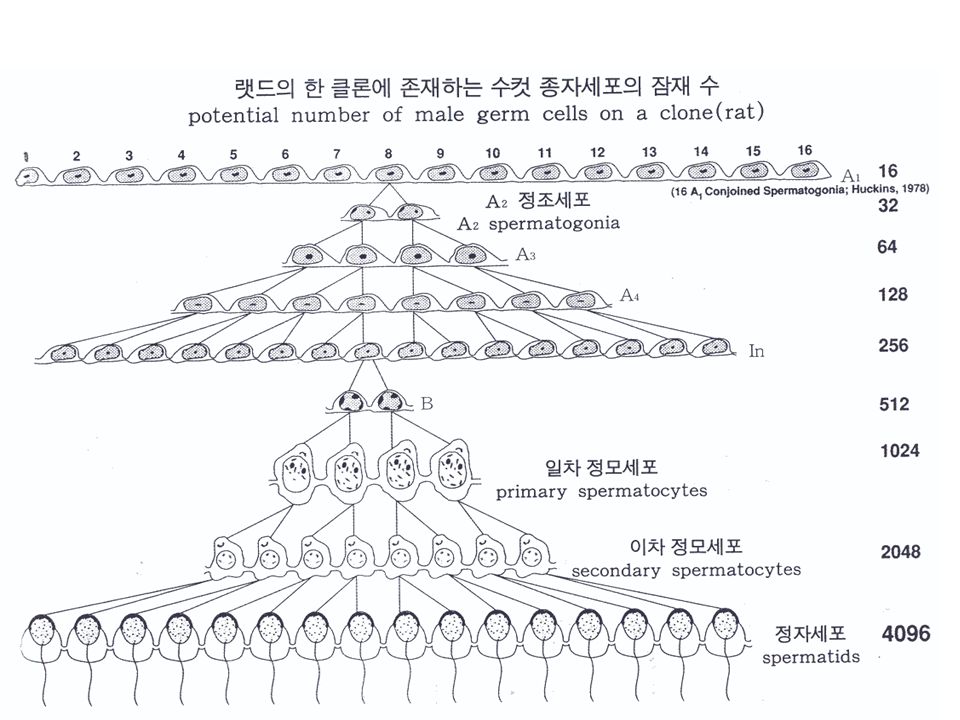
Testis (structure) Closely packed seminiferous tubules of 200~250 microns in diameter Human : separated by fascia ( 근막 ) Rodents : no subdivisions – Rat : 30 tubules / testis 1mm (length) / each tubule
 Closely packed seminiferous tubules of 200~250 microns in diameter Human : separated by fascia ( 근막 ) Rodents : no subdivisions – Rat : 30 tubules / testis 1mm (length) / each tubule")
4
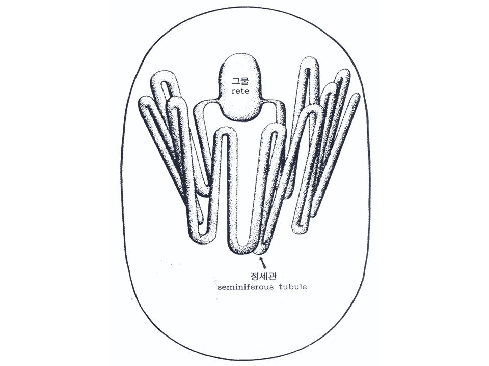
Testis (structure) Interstitium ( 정세관과 정세관 사이 ) –Blood and lymph vessels –Macrophages –Leydig cells (testosterone) Rete testis ( 정소망 ) –Ends of the seminiferous tubules –Intercommunicating system of channels lined with a low cuboidal/columnar epithelium –Upper pole of the testis (rat)
 Interstitium ( 정세관과 정세관 사이 ) –Blood and lymph vessels –Macrophages –Leydig cells (testosterone) Rete testis ( 정소망 ) –Ends of the seminiferous tubules –Intercommunicating system of channels lined with a low cuboidal/columnar epithelium –Upper pole of the testis (rat)")
9
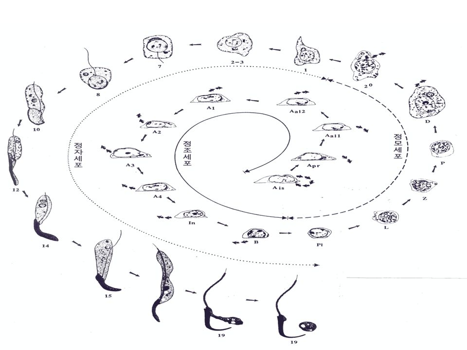
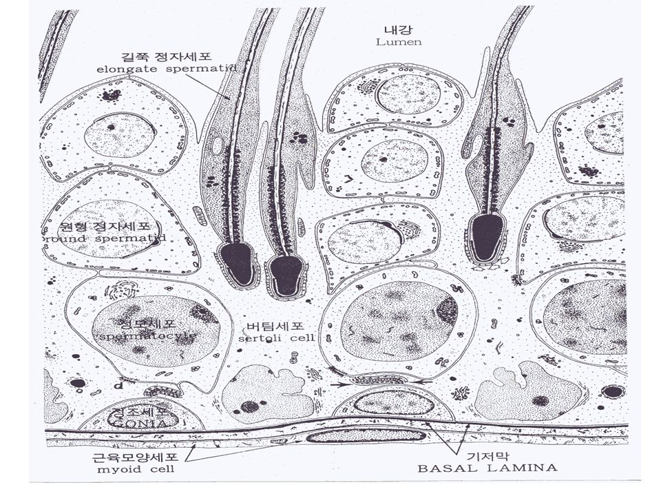
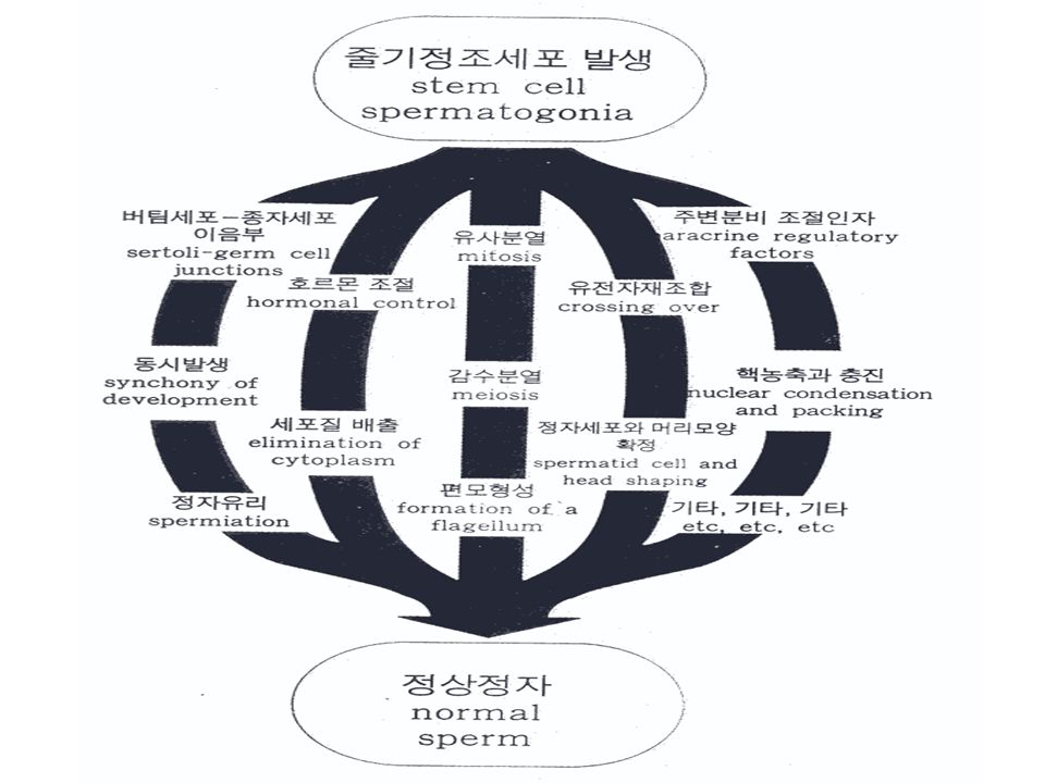
Gametogenesis( 배우자형성 ) The process of formation and development of gametes (germ cells) –Mitosis, meiosis and cell shape changes Seminiferous tubules –Sertoli cells –Developing germ cells Spermatogonia, primary spermatocytes, secondary spermatocytes, spermatid, spermatozoa –Myoid cells Aid the movement of spermatozoa and fluid along the lumen
 The process of formation and development of gametes (germ cells) –Mitosis, meiosis and cell shape changes Seminiferous tubules –Sertoli cells –Developing germ cells Spermatogonia, primary spermatocytes, secondary spermatocytes, spermatid, spermatozoa –Myoid cells Aid the movement of spermatozoa and fluid along the lumen")
10
Gametogenesis Spermatogenesis ( 정조세포 -> 정모세포 ) Spermiogenesis ( 정자세포 -> 정자 ) –Spermatids are reshaped into spermatozoa –Acrosome –Nuclear material condenses –A mid piece forms with aggregation of mitochondria –A flagellum is generated ( 편모형성 ) –Shedding of excess cytoplasm ( 허물 )
 Spermiogenesis ( 정자세포 -> 정자 ) –Spermatids are reshaped into spermatozoa –Acrosome –Nuclear material condenses –A mid piece forms with aggregation of mitochondria –A flagellum is generated ( 편모형성 ) –Shedding of excess cytoplasm ( 허물 )")
12
Sertoli cells Regulation of spermatogenesis and spermiogenesis `Nurse’ cells –Structural support –Provision of a specialized luminal fluid environment –Nutrition –Phagocytosis –Development and release of spermatozoa

14
Blood-testis barrier Sertoli-Sertoli cell barrier –Specialized luminal fluid environment –Protect spermatozoa from the immune system and harmful substances –Sertoli-Sertoli tight junctions –The barrier divides the seminiferous epithelium into 2 (basal and adluminal) compartment
 compartment")
16
The Epididymis Abandoned child of the reproductive system –Current : Motility and Morphology –Future : Sperm Proteins, Fertilization Assays Sperm Maturation in the Epididymis : Functional Change - Sperm acquire motility - Sperm acquire the ability to fertilize an oocyte Requires interaction between sperm, epididymal epithelium and luminal fluids

17
Origin of Adverse Effects on Epididymal Sperm Function Testicular Direct on epididymal spermatozoa Direct on epididymal epithelium Epididymal Toxicants (Few chemicals) Methyl Chloride, Alpha-chlorohydrin Alkane sulfonates
 Methyl Chloride, Alpha-chlorohydrin Alkane sulfonates")
18
Epididymis (Structure) Efferent ducts Initial segment Caput (head) Corpus (body) Cauda (tail) Distal Deferens
 Efferent ducts Initial segment Caput (head) Corpus (body) Cauda (tail) Distal Deferens")
19
Fertilization Spermatogenesis – Testis Maturation – Epididymis Fertilization – Female Reproductive Tract

20
Movement Fluid secreted by the Sertoli cells and the released spermatozoa pass along the lumen of the seminiferous tubule, into the rete testis, the efferent ducts and into the epididymis. Rate of movement within the lumen of a rat seminiferous tubule : 1 ㎕ /hour Myoid cells and Sertoli cells At this stage, spermatozoa are not able to swim.

21
Efferent ducts 6 efferent ducts join to form a single duct before entering into the epididymis (rat, mouse)
")
22
Efferent ducts Function –Reabsorption of water (90% of fluid leaving the testis) –Spermatozoa within the efferent ducts of the rat are concentrated (3%~20%) of the total volume
 –Spermatozoa within the efferent ducts of the rat are concentrated (3%~20%) of the total volume")
23
Epididymal duct Pseudostratified columnar epithelium Cells contain a brush border and large Golgi apparatus Surrounding the tubule is a muscular wall that thickens from proximal to distal regions.

24
Epididymal cell types Principal –Proximal resion > distal resion –Secretion and absorption of macromolecules, ions, organic solutes Narrow Clear –Absorption of macromolecules Basal –Protection of epithelium, spermatozoa from xenobiotics Halo –May be Monocytes or lymphocytes

25
Changes in the epithelium From proximal to distal regions –The height of epithelium is reduced –The luminal diameter increases –Appearance of different cell types –Presence or absence of stereocillia –Increase in the degree of smooth muscle surrounding the duct

26
Blood-Epididymis Barrier Among the various epithelial cell contents examined, the occlusion of the epididymis is the most highly developed. Tight junction separate the basal and luminal spaces.

27
Luminal Fluid Microenvironment The composition changes dramatically from proximal to distal regions. The composition is the result of secretion and absorption of molecules across the epididymal epithelium. Changing environment is responsible for changes that occur within spermatozoa as they progress along the epididymal duct

28
Sperm Maturation Spermatozoon (pl. spermatozoa) The relative position along the epididymal duct for varies species where spermatozoa acquire the ability to fertilize an egg. –rabbit (corpus) –mouse, rat, man (cauda)
 The relative position along the epididymal duct for varies species where spermatozoa acquire the ability to fertilize an egg. –rabbit (corpus) –mouse, rat, man (cauda).")
29
Changes in Spermatozoa During Epididymal Transit Surface charge Lectin-binding properties Nuclear bonds Lipid and carbohydrate composition Membrane fluidity Surface proteins Many of these changes continue through ejaculation, acrosome reaction, fertilization

30
Sperm transport (days) 5 10 15 Monkey Rabbit Rat Man
 Monkey Rabbit Rat Man")
31
Sperm Storage Sp. In Cauda (x 1,000,000) / Daily Sp. Production Man 3 Rat 5 Rabbit 10
 / Daily Sp. Production Man 3 Rat 5 Rabbit 10")
32
Sperm Protection Spermatozoa must be protected from the immune system, harmful xenobiotics and agents that may cause oxidative stress. The blood-epididymis barrier plays a major role in the protection of sperm.

33
정소독성평가 1) 정소 육안관찰 ( 장기무게, atrophy) 2) 정세관 전체 관찰 (tubular necrosis) 3) 정세관내 생식세포 관찰 (spermatogenesis) 4)Sertoli cells 관찰 5)Leydig cells 관찰 6) 정소상체 : caput, corpus, cauda 관찰 –Aberrant cell types in lumen –Absence of clear cells in cauda
 정소 육안관찰 ( 장기무게, atrophy) 2) 정세관 전체 관찰 (tubular necrosis) 3) 정세관내 생식세포 관찰 (spermatogenesis) 4)Sertoli cells 관찰 5)Leydig cells 관찰 6) 정소상체 : caput, corpus, cauda 관찰 –Aberrant cell types in lumen –Absence of clear cells in cauda")
34
Germ cells Assesment of testicular degeneration (a) Subjective assessment using modifiers - mild, moderate, severe (b) Measurement of the frequency of abnormal tubules (c) No. of XIV stage / No. of the total tubules (d) Selected degrees of tubular abnormality severe degrees = Classify the eight grades of degeneration x No. of tubules (e) Sertoli cell index (SCI) SCI = No. of germinal cells / No. of Sertoli cells
 Selected degrees of tubular abnormality severe degrees = Classify the eight grades of degeneration x No. of tubules (e) Sertoli cell index (SCI) SCI = No. of germinal cells / No. of Sertoli cells.")
35
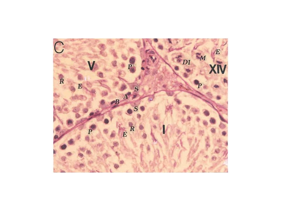
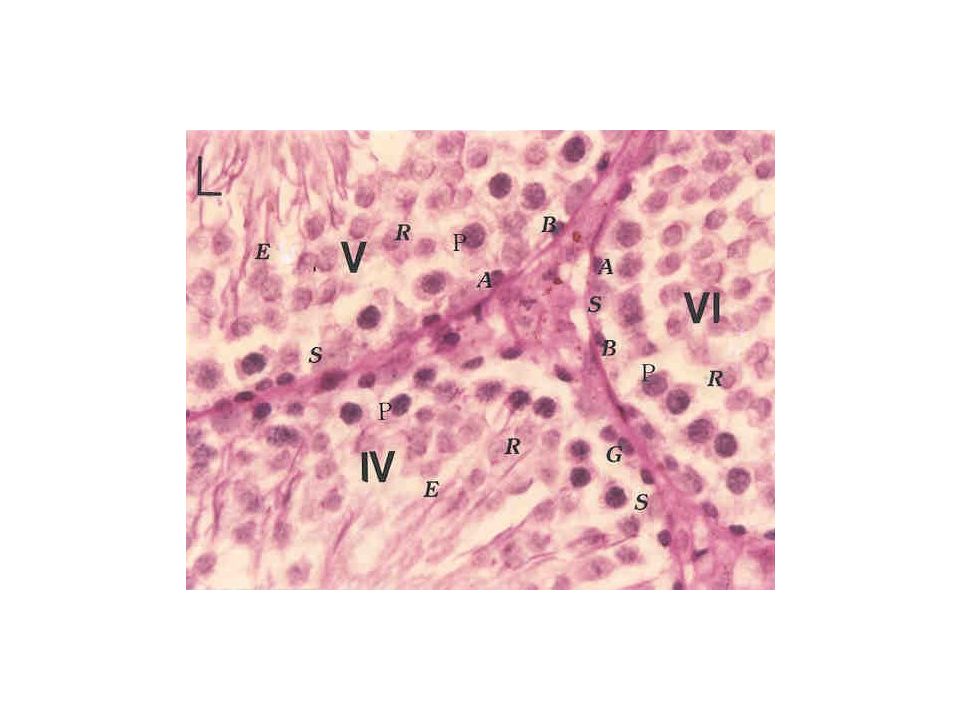
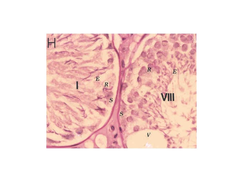
정조, 정모세포 stage II-III 정조세포의 괴사 Sertoli cell 의 탐식작용 stage VII – ( 정조세포, preleptotene, pachytene, 정자세포 ) 정조세포, preleptotene 정모세포 소실 정세관 기저층에 pachytene 정모세포
 정조세포, preleptotene 정모세포 소실 정세관 기저층에 pachytene 정모세포")
36
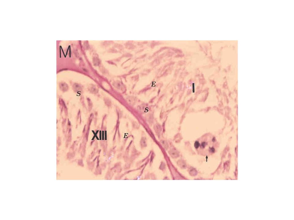
정자세포 Stage IX Sertoli cell 의 장해 (* 공포형성 ) 정세관의 기저부에 정자세포 (step 19) 가 박리되 지 않고 잔류 되어 있다. Stage VI 정자세포의 다핵거대화 Sertoli cell 장해에 따른 변화

37
Tubular necrosis Tubular necrosis occurs as a results of ischemia Cadmium (testicular blood supply) - cadmium 단회 투여 후 2 일차 (rat) - 혈관내피 장해 - ischemia ( 허혈 ), 순환장해 - 정소의 경색상 변화 - cadmium 단회 투여 후 7 일차 (rat) - 정세관 괴사 - 간질의 염증 반응 - stage 특이성, 세포 특이성이 보이지 않는다.
 - cadmium 단회 투여 후 2 일차 (rat) - 혈관내피 장해 - ischemia ( 허혈 ), 순환장해 - 정소의 경색상 변화 - cadmium 단회 투여 후 7 일차 (rat) - 정세관 괴사 - 간질의 염증 반응 - stage 특이성, 세포 특이성이 보이지 않는다.")
38
Sertoli cells Sertoli cells do not divided after 18-21 days of age. Structural support and movement of germ cells. * 19 spermatid retention at VIII stage Phagocytic activity. Formation of the blood-tubule barrier Cellular metabolism and intercellular interactions Sertoli-only syndrome –Ethanol 단회 투여 후 14 일차 (rat) – 생식세포의 소실
 – 생식세포의 소실.")
39
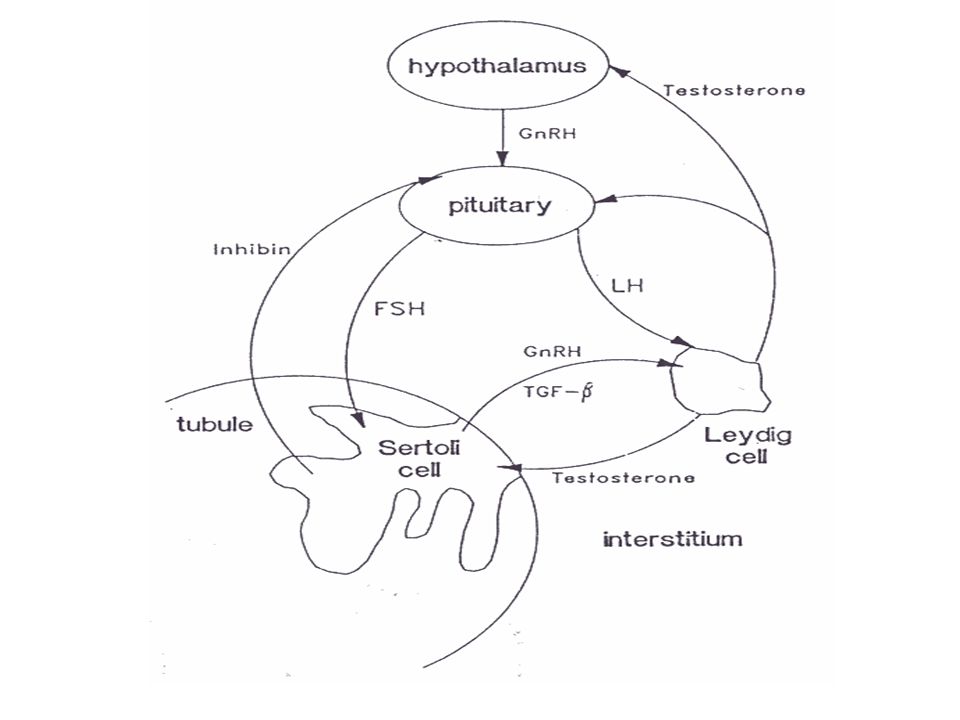
Leydig cells - The major site for the synthesis of testosterone. -Large pale cells in the pituitary known as castration cells are often found in association with severe bilateral testicular atrophy and Leydig cell hyperplasia. - These are presumed to secrete gonadotropic hormones.

40
Leydig cell loss Ethanol 단회 투여 후 7 일차 (rat) Leydig cell 소실 Testosterone 생산저하 정자세포, 정모세포 의 변성 및 괴사
 Leydig cell 소실 Testosterone 생산저하 정자세포, 정모세포 의 변성 및 괴사")
41
Leydig cell hyperplasia Leydig cells 증가 Pituitary 내의 castration cells 출현 –Leydig cell 의 testosterone 분비 기능조절은 pituitary 에서 분비되는 LH 에 의하여 조절된다. –Testosterone levels 은 pituitary 의 LH 분비를 조절한다. Leydig cell 의 testosterone 분비기능 이상은 pituitary 의 LH 분비를 자극하여 Leydig cell hyperplasia 를 초래한다.

42
Finding of primary target cell -Time course study of spermatogenesis -Maturation depletion : The progressive loss of subsequent cell types following injury to a precursor cell

43
표적세포 투여기간, 투여농도 에 따라 표적세포는 달라질 수 있다. 생식세포는 세르토리세포 에 의하여 24 시간 내에 탐 식된다. 정자세포는 정세관 내강 으로 계속 빠져나가므로 표적 세포를 찾는데 혼란스러울 수 있다. 동일한 스테이지 의 각 생식세포의 종류별 수 비교 통계적 유의성 (SCI) : 표적세포
 : 표적세포.")
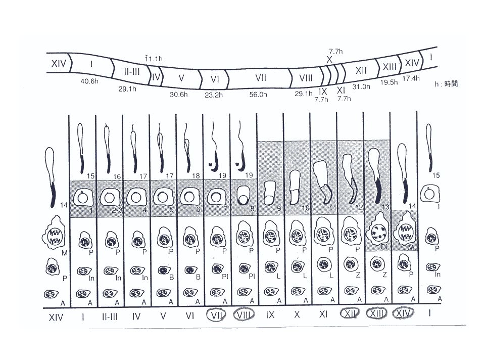
44
정자형성과정 - 14 different stages of cellular association : 19 different steps of spermatid (acrosome structure) - Rat : 48-53 days (a cycle time of 12-13.3 days) - 스테이지 별로 소요되는 시간 : 각 스테이지마다 다르며 정소에서 출현하는 정세관 스테이지 빈도수와 비례하게 된다 (Haschek and Rousseaux 1991).
 - Rat : days (a cycle time of days) - 스테이지 별로 소요되는 시간 : 각 스테이지마다 다르며 정소에서 출현하는 정세관 스테이지 빈도수와 비례하게 된다 (Haschek and Rousseaux 1991).")
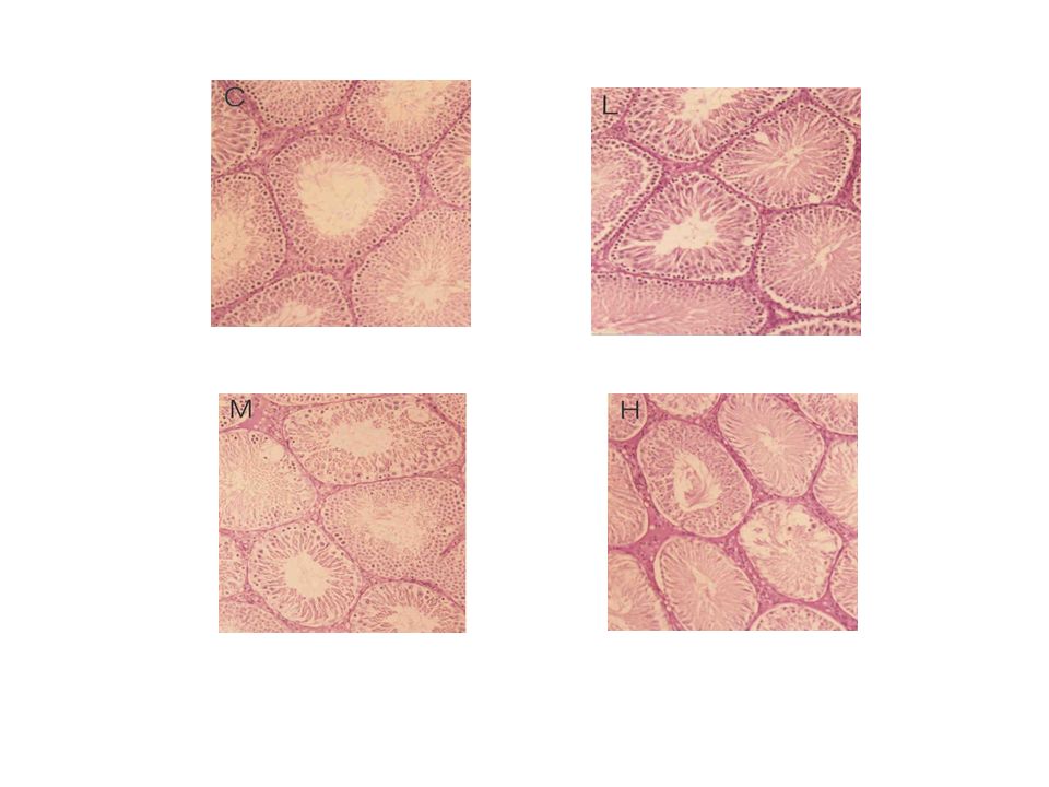
46
Table 1. Body weight (g) and relative testes weight (mg) of male rats treated with 2- bromopropane for 28 days Control125 mg/kg250 mg/kg500 mg/kg Initial body weight 300.5 ± 14.3300.7 ± 12.7300.3 ± 12.7301.4 ± 13.2 Terminal body weight 356.7 ± 16.1352.5 ± 11.8 330.4 ± 10.7* 289.8 ± 16.0* Testes (L)475.5 ± 30.7467.9 ± 36.7 402.9 ± 49.3* 329.8 ± 88.8* Values are means ± S.D. Significantly different from control group at * p<0.05 or ** p<0.01. Relative testes weight = (testes weight / terminal body weight) x 100g. L: Left.
 and relative testes weight (mg) of male rats treated with 2- bromopropane for 28 days Control125 mg/kg250 mg/kg500 mg/kg Initial body weight ± ± ± ± 13.2 Terminal body weight ± ± ± 10.7* ± 16.0* Testes (L)475.5 ± ± ± 49.3* ± 88.8* Values are means ± S.D. Significantly different from control group at * p<0.05 or ** p<0.01. Relative testes weight = (testes weight / terminal body weight) x 100g. L: Left..")
48
Table 2. Numbers of Sertoli cells and Sertoli cells indices (SCI) of rats treated with 2-bromopropane for 28 days Control125 mg/kg250 mg/kg500 mg/kg No. of Sertoli cells 17.7 ± 2.216.8 ± 1.213.6 ± 1.0*16.1 ± 1.8 SCI30.2 ± 2.826.9 ± 4.7 21.0 ± 3.0** 11.8 ± 1.4** Values are means ± S.D. Significantly different from control group at * p<0.05 or ** p<0.01. SCI = Total number of germ cells / total number of Sertoli cells. Total numbers of testes in each group = 6. Number of tubules in each testis = 13.
 of rats treated with 2-bromopropane for 28 days Control125 mg/kg250 mg/kg500 mg/kg No. of Sertoli cells 17.7 ± ± ± 1.0*16.1 ± 1.8 SCI30.2 ± ± ± 3.0** 11.8 ± 1.4** Values are means ± S.D. Significantly different from control group at * p<0.05 or ** p<0.01. SCI = Total number of germ cells / total number of Sertoli cells. Total numbers of testes in each group = 6. Number of tubules in each testis = 13..")
53
Table 3. Sertoli cells indices (SCI) of each spermatogenic cell of rats treated with 2- bromopropane for 28 days Control125 mg/kg250 mg/kg500 mg/kg SCI Spermatogonia 1.2 ± 0.1 1.0 ± 0.1* 0.4 ± 0.2** 0.3 ± 0.1** Spermatocyte preleptotene3.6 ± 0.6 2.6 ± 0.5** 1.0 ± 0.2** 0.0 ± 0.0** leptotene 4.1 ± 0.73.2 ± 0.5 1.6 ± 0.2** 0.2 ± 0.1** zygotene 4.6 ± 0.73.9 ± 0.6 1.5 ± 0.6** 0.0 ± 0.0** pachytene4.7 ± 0.6 3.8 ± 0.3** 2.0 ± 0.9** 0.0 ± 0.0** diplotene 5.9 ± 1.15.9 ± 1.2 2.5 ± 2.1* 0.0 ± 0.0** Spermatid round 15.1 ± 1.4 12.7 ± 0.7* 9.8 ± 2.2** 1.8 ± 0.6** elongate 12.1 ± 1.111.0 ± 1.012.1 ± 0.910.4 ± 1.4* Values are means ± S.D. Significantly different from control group at * p<0.05 or ** p<0.01. SCI = each number of germ cells / total number of Sertoli cells. Total numbers of testes in each group = 6. Number of tubules in each testis = 13.
 of each spermatogenic cell of rats treated with 2- bromopropane for 28 days Control125 mg/kg250 mg/kg500 mg/kg SCI Spermatogonia 1.2 ± ± 0.1* 0.4 ± 0.2** 0.3 ± 0.1** Spermatocyte preleptotene3.6 ± ± 0.5** 1.0 ± 0.2** 0.0 ± 0.0** leptotene 4.1 ± ± ± 0.2** 0.2 ± 0.1** zygotene 4.6 ± ± ± 0.6** 0.0 ± 0.0** pachytene4.7 ± ± 0.3** 2.0 ± 0.9** 0.0 ± 0.0** diplotene 5.9 ± ± ± 2.1* 0.0 ± 0.0** Spermatid round 15.1 ± ± 0.7* 9.8 ± 2.2** 1.8 ± 0.6** elongate 12.1 ± ± ± ± 1.4* Values are means ± S.D. Significantly different from control group at * p<0.05 or ** p<0.01. SCI = each number of germ cells / total number of Sertoli cells. Total numbers of testes in each group = 6. Number of tubules in each testis = 13..")
54
Table 4. At indicated stage, Sertoi cell indices (SCI) % of each germ cell of rats treated with 2-Bromopropane 125 mg/kg for 28 days stagespermatogonia spermatocytes spermatids preleptoteneleptotenezygotenepachytenediploteneround elongate I100±25 79±20 87±3494±43 II-III54±11** 92±44 94±44100±37 IV108±31 99±26 99±23102±17 V83±40 78±16* 84±2890±30 VI78±9** 85±11* 89±10*94±25 VII104±5368±28 83±44 72±2556±22 VIII90±3056±35* 75±29 77±2982±33 IX70±45 89±28 88±36 104±41 X50±18** 73±27* 80±27 98±13 XI66±27* 86±18 101±30 89±27 XII112±37 82±14*92±21 95±22 XIII66±19* 87±28 102±30 104±20 XIV 95±59 83±35103±38 98±29 Values are means ± S.D. Significantly different from control group at * p<0.05 or ** p<0.01. SCI = The number of the each germinal cells / total number of Sertoli cells. Date are expressed as % of control value. Total numbers of testes = 6. Number of tubules in each testis = 13.
 % of each germ cell of rats treated with 2-Bromopropane 125 mg/kg for 28 days stagespermatogonia spermatocytes spermatids preleptoteneleptotenezygotenepachytenediploteneround elongate I100±25 79±20 87±3494±43 II-III54±11** 92±44 94±44100±37 IV108±31 99±26 99±23102±17 V83±40 78±16* 84±2890±30 VI78±9** 85±11* 89±10*94±25 VII104±5368±28 83±44 72±2556±22 VIII90±3056±35* 75±29 77±2982±33 IX70±45 89±28 88±36 104±41 X50±18** 73±27* 80±27 98±13 XI66±27* 86±18 101±30 89±27 XII112±37 82±14*92±21 95±22 XIII66±19* 87±28 102±30 104±20 XIV 95±59 83±35103±38 98±29 Values are means ± S.D. Significantly different from control group at * p<0.05 or ** p<0.01. SCI = The number of the each germinal cells / total number of Sertoli cells. Date are expressed as % of control value. Total numbers of testes = 6. Number of tubules in each testis = 13..")
55
표적세포 spermatogonia, spermatocytes, 초기 spermatids 에서 대 조군에 비하여 통계적으로 유의한 생식세포의 감소. 시험물질이 spermatogonia 에 영향을 미쳐 4 주 후, spermatogonia, spermatocytes, 초기 spermatids 의 손실 로 나타난 것이다. 만일 spermatocytes 에 영향을 미쳤다면, spermatocytes 와 후기 spermatids 의 감소가 나타났을 것이고, spermatogonia 의 수는 변화가 없었을 것이다. 결론적으로 정자발생과정과 세르토리세포의 탐식작용을 고려할 때, 시험물질의 표적세포는 spermatogonia.

56
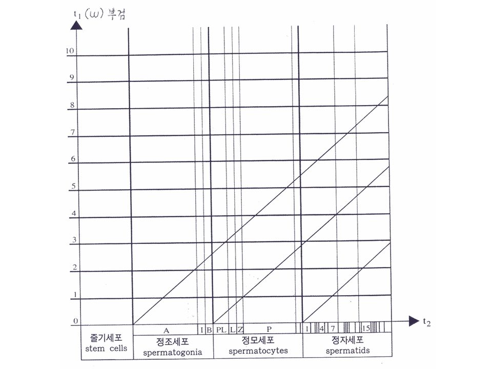
정소독성변화의 해석 - 생식세포중 표적세포의 추정 (Ettlin, 1984) - 단회 투여 (rat) - 종축은 투여 후 주, 횡축은 각종 생식세포 - 종축의 각종 생식세포의 간격은 정자형성 cycle (spermatogenesis) 진행시간을 고려한 것임. 예 : 단회투여 후, 3 주 후에 부검하여 zygotene 정모세포 감소 * 투여 후 3 주 의 횡축의 zyrotene 정모세포의 위치에 점을 찍고, * 챠트의 사선과 평행한 선을 아래쪽으로 그어 보면, * 그 선의 선단은 정조세포 type A 와 만난다. * 이 세포가 표적세포로 추정된다.

58
표본제작 1. 고정 : Bouin 용액에 고정 ( 약 24 시간 이내 ) 1) 2-3 시간 고정 2) 침투를 양호하게 한 후 가운데 부근에 횡단할면을 넣어 재고정 3) 약 12 시간 후 3-4mm 의 두께로 절취, 재고정 2. 절취 : 1) 좌우의 정소를 횡단하고 3-4mm 두께 2) 정소상체 : 종 (longitudinal section) 절취 3. 조직처리 4. 포매 5. 박절
 1) 2-3 시간 고정 2) 침투를 양호하게 한 후 가운데 부근에 횡단할면을 넣어 재고정 3) 약 12 시간 후 3-4mm 의 두께로 절취, 재고정 2. 절취 : 1) 좌우의 정소를 횡단하고 3-4mm 두께 2) 정소상체 : 종 (longitudinal section) 절취 3. 조직처리 4. 포매 5. 박절.")
59
6. 염색 : PAS 1) 탈파라핀, 함수 2) 0.5% Periodic acid 20 분 3) D.W 수세 4) Schiff reagent 30 분 5) 흐르는 물에 수세 6) Hematoxylin 대조염색 15 분 7) 흐르는 물에 수세 8) 탈수 9) 투명 10) 봉입
 탈파라핀, 함수 2) 0.5% Periodic acid 20 분 3) D.W 수세 4) Schiff reagent 30 분 5) 흐르는 물에 수세 6) Hematoxylin 대조염색 15 분 7) 흐르는 물에 수세 8) 탈수 9) 투명 10) 봉입.")
Similar presentations


. Why must organisms reproduce?>")

 2008 년 10 월 ( 사단법인 ) 사회적기업 청람 Co. social enterprise cheong ram 영광종합병원 · 공립영광노인전문요양병원 의료법인 호연재단.>")

 사업명 (HY 중고딕 20) 사업위치 (HY 중고딕 20) 민간건축물 미작성.>")

 Ch 10 Reproductive system.>")


 비만률순위 : 29 위 (2005 OECD)>")