Download presentation
Presentation is loading. Please wait.

1
KCP 790 연세대학교 의과대학 세브란스 병원 전공의 오은지

2
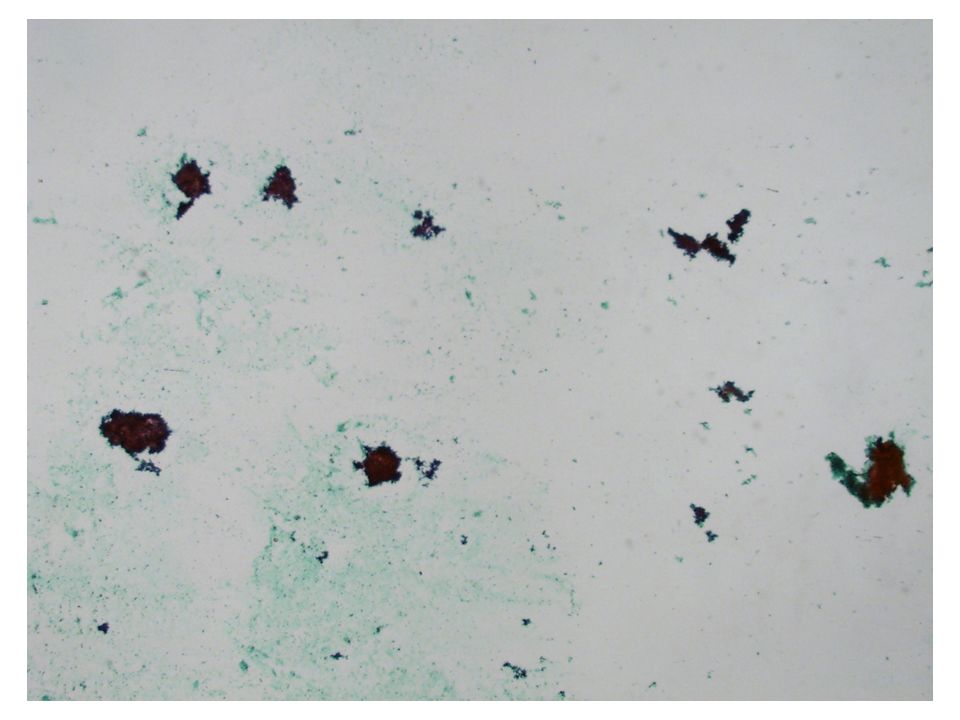
Patient history M/64 특이병력 없음 어지러움을 주소로 내원 영상 소견 – 뇌 MRI, 흉부 CT 상에서 다발성 뇌 경색 및 폐 색전증 소견 보였으 며, 경부 CT 상 다발성의 좌측 level Ⅲ, Ⅳ, Ⅴ, 쇄골상 림프절 비대가 관찰되었고, 복부 CT 상에서도 양측 서혜부, 외장골, 내장골, 총장 골, 대동맥 주위 림프절 비대 소견이 관찰되었습니다. 좌측 쇄골상 림프절에서 세침흡인

7
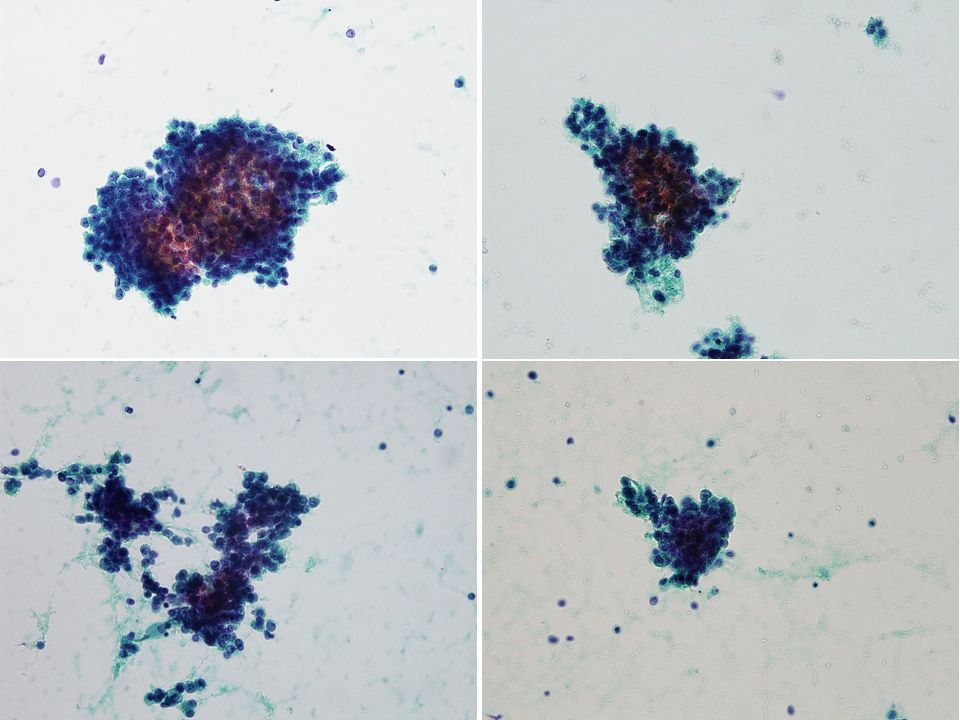
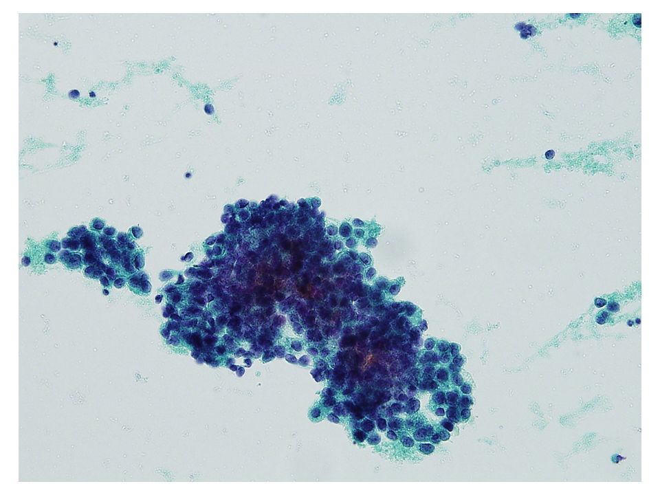
Cytologic findings Lymphoid background Cell clusters: papillary, solid, acinar, and microacinar arrangement Single scattered cells Small uniform round cells Overlapping High N/C ratio Hyperchromatic Prominent nucleoli Delicate, fine and focal vacuolated cytoplasm

8
Differential diagnosis Lymphoma Metastatic carcinoma – Well differentiated adenocarcinoma Biliary tract/stomach/colon/lung etc. – Prostatic adenocarcinoma – Breast lobular carcinoma – Neuroendocrine tumor

9
Lymphoma – Monotonous, equal size 의 lymphoid cell – Cluster 를 이루지 않고 single cell 로 나옴 Well differentiated adenocarcinoma (biliary tract/lung/stomach/colon etc.) – Many individual cells + 3-D clusters – Cohesiveness↓, polarity↓, nuclear enlargement, high N/C ratio, prominent nucleoli, hyperchromasia, variable mucin – Biliary tract: Large 3-D cluster, coarse chromatin clumping – Lung: 3-D cluster (acini, tubule, papillae), fine granular to powdery chromatin, round to oval nuclei, variable cell size
 – Many individual cells + 3-D clusters – Cohesiveness↓, polarity↓, nuclear enlargement, high N/C ratio, prominent nucleoli, hyperchromasia, variable mucin – Biliary tract: Large 3-D cluster, coarse chromatin clumping – Lung: 3-D cluster (acini, tubule, papillae), fine granular to powdery chromatin, round to oval nuclei, variable cell size")
10
Neuroendicrine tumor 1) Carcinoid Dispersed or small groups (forming flat, loosely structured gland like clusters) Uniform appearance: cuboidal or rectangular with faintly basophilic transparent cytoplasm and eccentric nuclei, plasmacytoid nuclei, fine granular chromatin (“salt and pepper”), tiny nucleoli, occasional giant cells 2) Well differentiated neuroendocrine carcinoma (atypical carcinoid) Organoid arrangement, high mitotic rate, prominent nucleoli (single or multiple) and focal necrosis variable nuclear size, hyperchromasia, abundant cytoplasm, non- pyknotic ovoid nuclei, clear cytoplasm
 Carcinoid Dispersed or small groups (forming flat, loosely structured gland like clusters) Uniform appearance: cuboidal or rectangular with faintly basophilic transparent cytoplasm and eccentric nuclei, plasmacytoid nuclei, fine granular chromatin ( salt and pepper ), tiny nucleoli, occasional giant cells 2) Well differentiated neuroendocrine carcinoma (atypical carcinoid) Organoid arrangement, high mitotic rate, prominent nucleoli (single or multiple) and focal necrosis variable nuclear size, hyperchromasia, abundant cytoplasm, non- pyknotic ovoid nuclei, clear cytoplasm")
11
Breast lobular carcinoma – Small cell size, minimal atypia, intracytoplasmic vacuole – Indistinct nucleoli, intracytoplasmic neolumina (12-57%) – Dispersed cells, no acini – Extremely rare (1% of male breast cancer) Prostatic adenocarcinoma – 비교적 uniform 한 cell 들의 small cluster or acini – Small, round to oval cells – Enlarged, round nuclei, increased N/C ratio, moderate amount of cytoplasm – Microacinar pattern 과 prominent nucleoli 가 가장 특징적인 소견
 – Dispersed cells, no acini – Extremely rare (1% of male breast cancer) Prostatic adenocarcinoma – 비교적 uniform 한 cell 들의 small cluster or acini – Small, round to oval cells – Enlarged, round nuclei, increased N/C ratio, moderate amount of cytoplasm – Microacinar pattern 과 prominent nucleoli 가 가장 특징적인 소견")
12
Differential diagnosis Lymphoma Metastatic carcinoma – Well differentiated carcinoma Biliary tract/stomach/colon/lung etc. – Prostatic adenocarcinoma – Breast lobular carcinoma – Neuroendocrine tumor

13
2007 2011 Diagnostic Cytopathology

14
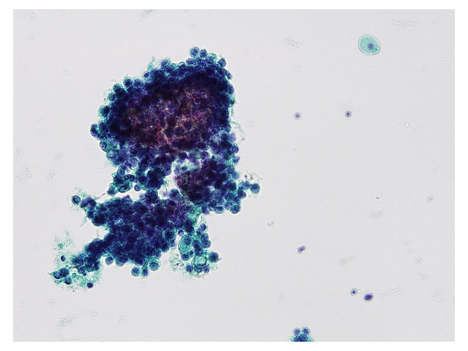
Small- to medium sized relatively uniform cells Small clusters forming acini, solid, and cirbriform microacinar + scattered single cells Loss of honeycomb Moderately enlarged, hyperchromatic nuclei with mild nuclear membrane irregularities Nuclear membrane irregularity, hyperchromasia 와 chromatin clumping 은 다양하게 나타나지만 often minimal Prominent nucleoli (single or multiple) Delicate and finely vacuolated cytoplasm Neuroendocrine differentiation (10-33%) Immunohistochemical pannel – PSA (prostate-specific antigen), PSAP (prostatic acid phosphatase), and AMACR (alpha- methylacyl-CoA racemase)
 Delicate and finely vacuolated cytoplasm Neuroendocrine differentiation (10-33%) Immunohistochemical pannel – PSA (prostate-specific antigen), PSAP (prostatic acid phosphatase), and AMACR (alpha- methylacyl-CoA racemase)")
15
Diagnosis Positive for malignancy, metastatic carcinoma, see note. Note) 1.The possibility of metastatic adenocarcinoma from prostate can be suggested. 2.Further evaluation is recommended.
 1.The possibility of metastatic adenocarcinoma from prostate can be suggested. 2.Further evaluation is recommended..")
16
감사합니다.

Similar presentations
. CC: Low abd distension for 1 M CC: Low abd distension for 1 M (Pap: N these day, CA125: 15) (Pap: N these day, CA125: 15) Wt loss.>")

 제출자 발표 2012.02.01 서울성모병원 병리과 전공의 이영섭.>")

 2) 3) 4) 2. 1) 2) 3. 1) 2) 3) * 혈액의 생성과 파괴 혈구생성부위 -- 1. 2. 3. * 조혈자극인자 –- 1. 2. 3. * 적혈구의 분화와 성숙과정 – Pluripotent stem cell →>")


 강의실 9호관 506호 보건행정학과 2학년 B,C반.>")


