Download presentation
Presentation is loading. Please wait.

1
Persistent Hyperplastic Primary Vitreous
<망막 conference> Persistent Hyperplastic Primary Vitreous R3 김나현/St. 신정아/AP.박영훈

2
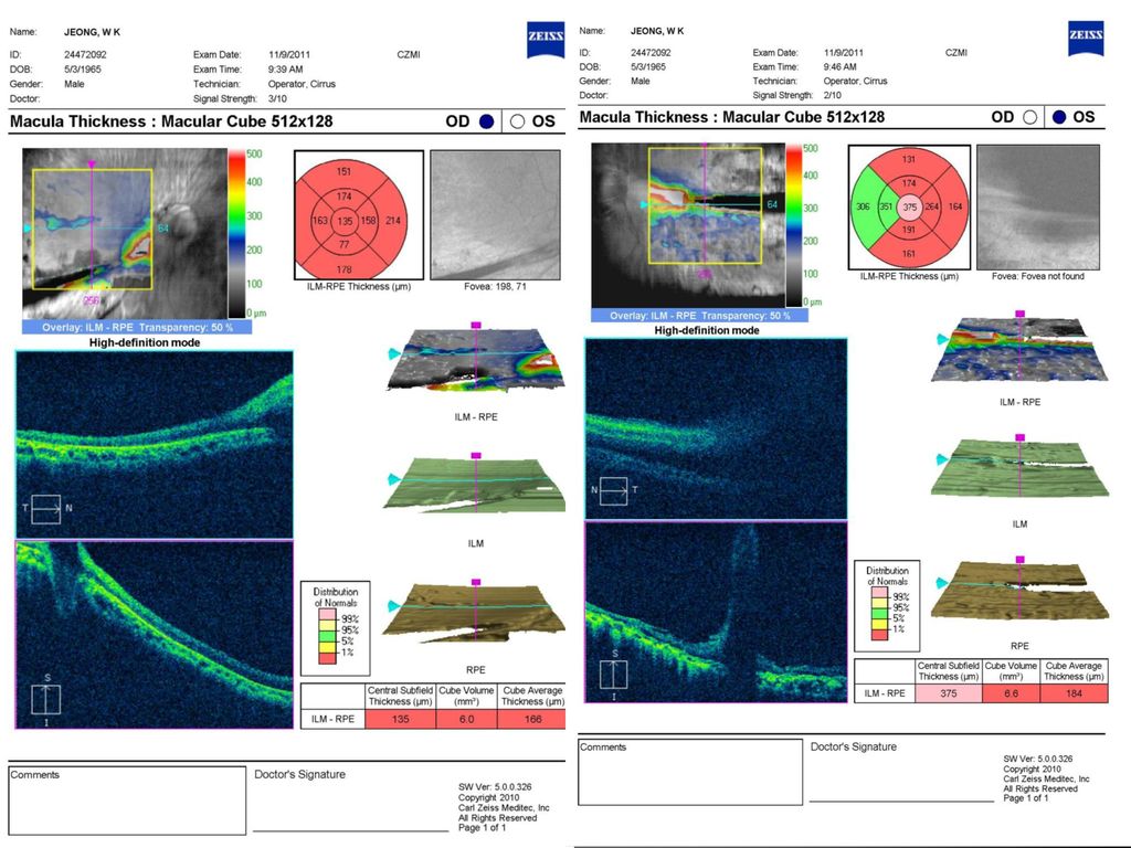
Case 1. 24472092 48/M 정 O 교 C.C ) “물체가 작게 보이고 흐려 보여요”(OS)
onset) For 3 months 상환 어렸을적 선천적 망막 이상 있다고 들었던 분으로 내원 3개월 전 좌안 백내장 수술 받은 후부터 물체가 작게 보이고 흐려 보이는 증상 있어 내원 DM/HBP (-/-) Past Hx : 어렸을적 선천적 망막 이상 있다고 들음 Ocular op/trauma(+/-) : phaco+PCL(OS) at 강릉아산병원 Gls (-) Eye drop (-)
 For 3 months. 상환 어렸을적 선천적 망막 이상 있다고 들었던 분으로 내원 3개월. 전 좌안 백내장 수술 받은 후부터 물체가 작게 보이고 흐려 보이는. 증상 있어 내원. DM/HBP (-/-) Past Hx : 어렸을적 선천적 망막 이상 있다고 들음. Ocular op/trauma(+/-) : phaco+PCL(OS) at 강릉아산병원. Gls (-) Eye drop (-)")
3
Ocular examination VA OD 0.02 (N-C x +0.25Ds : -9.50Dc Ax64)
OS FC/50cm (N-C) IOP OD/OS 17/9 mmHg EOM XT=30PD by H-test, no LOM(OU) Conj. OU not injected Cornea OU clear AC OU deep & cell (-) Pupil OU round & nl sized, LR(+/+) Lens OD mild cortical opacity, PSCO OS PCL in situ c mild PCO(+) Fds OD disc dragging to inferotemporal area, linear vitreous traction line, Retinal atrophy , pigmented lesion in inferotemporal area OS disc dragging to inferotemporal area, linear vitreous traction line, Retinal atrophy , pigmented lesion in inferotemporal area
 IOP OD/OS 17/9 mmHg. EOM XT=30PD by H-test, no LOM(OU) Conj. OU not injected. Cornea OU clear. AC OU deep & cell (-) Pupil OU round & nl sized, LR(+/+) Lens OD mild cortical opacity, PSCO. OS PCL in situ c mild PCO(+) Fds OD disc dragging to inferotemporal area, linear vitreous traction. line, Retinal atrophy , pigmented lesion in inferotemporal area. OS disc dragging to inferotemporal area, linear vitreous traction line, Retinal atrophy , pigmented lesion in inferotemporal area.")
4
Impression PHPV(OU) Plan FAG M-OCT 360’ fundus photo
 Plan FAG M-OCT 360’ fundus photo")
5
FAG

9
F/U Because of Localized RD (OS),
barrier laser(OS) by AP.박영훈 add. barrier laser(OS) by AP.박영훈 were done.
 by AP.박영훈 add. barrier laser(OS) by AP.박영훈 were done.")
10
Case 2. 23797993 41/M 양 O 호 C.C ) Dec.VA (OD)
상환 좌안은 어릴 때부터 광각무 상태였던 분으로 내원 2주전부터 갑자기 지속된 우안 통증 및 시력 저하로 local 안과 내원하여 R/O ACG(OD) 의심하에 mannitolization 및 안압약 점안 이후 증상은 완화되었으며 내원 1주전 울산대 병원 내원하여 시행한 검사상 B scan 상 망막박리 의심되어 본원 내원함. DM/HBP (-/-) Ocular op/trauma(-/-) Gls (-) Eye drop (+) : O-LON X 4, CRAVIT X 4, O-2HOM X 4 /OD - 현재 안압 하강제 점안 안함 FHx : n-s
 의심하에. mannitolization 및 안압약 점안 이후 증상은 완화되었으며 내원 1주전. 울산대 병원 내원하여 시행한 검사상 B scan 상 망막박리 의심되어 본원 내원함. DM/HBP (-/-) Ocular op/trauma(-/-) Gls (-) Eye drop (+) : O-LON X 4, CRAVIT X 4, O-2HOM X 4 /OD. - 현재 안압 하강제 점안 안함. FHx : n-s.")
11
Ocular examination B scan : R/O RD (OD) VA OD HM (N-C) OS LP(-) (N-C)
IOP OD 45 OS <5 mmHg at 9:35 am Lid OU No swelling Conj. OU not injected Cornea OD sl. edematous c diffuse SPE c microbullae(+) OS clear AC OD 2.5CT at center, 1/4CT at pph. & cell(-) OS >4CT at center, >1/2CT at pph. & cell(-) Pupil OD round & mod dilated state OS round & miotic state, 360' post synechiae Lens OD mature cataract OS invisible Fd OD blurry invisible d/t cataract B scan : R/O RD (OD)
 OS clear. AC OD 2.5CT at center, 1/4CT at pph. & cell(-) OS >4CT at center, >1/2CT at pph. & cell(-) Pupil OD round & mod dilated state. OS round & miotic state, 360 post synechiae. Lens OD mature cataract. OS invisible. Fd OD blurry invisible d/t cataract. B scan : R/O RD (OD)")
12
Impression Plan Intumescent cataract(OD) R/O Phacomorphic glaucoma(OD)
R/O RD(OD) Prephthisis bulbi(OS) Plan Phaco + ppV (OD)
 Prephthisis bulbi(OS) Plan. Phaco + ppV (OD)")
13
Op. record Diagnosis : PHPV c total RD c PVR(OD) Mature cat(OD)
Op. title : ppV+ppL+MP+FAE+PFCL inj.+ removal+endolaser+S.encircling+SO inj.(OD) Op. finding: PHPV(+) (stalk arising from disc, total RD)
 Op. finding: PHPV(+) (stalk arising from disc, total RD)")
14
F/U <POD#3d> <POD #3d> VA : HM IOP(AP) 14 mmHg at 7:00am
(O-COST x 2, O-BMDP x 2, O-TVT x hs /OD) Conj. well-approximated wx. Cornea mild edematous c 3x4 mmsized epiedefect at center AC deep & cell(+++, RBC), SO bubble(+) Pupil ovoid & mild dilated d/t cycloplegics Lens A. aphakia Fds Glistering app. d/t SO, stalk arising from disc, seems to be flat post.pole, well-applied laser scars at 5 o/c, well elevated encircling band /OD
 Conj. well-approximated wx. Cornea mild edematous c 3x4 mmsized epiedefect at center. AC deep & cell(+++, RBC), SO bubble(+) Pupil ovoid & mild dilated d/t cycloplegics. Lens A. aphakia. Fds Glistering app. d/t SO, stalk arising from disc, seems to be flat. post.pole, well-applied laser scars at 5 o/c, well elevated encircling band. /OD.")
15
POD#8m VA OS HM

16
Persistent Hyperplastic Primary Vitreous
<Review> Persistent Hyperplastic Primary Vitreous

17
Definition Congenital anomaly of the eye that results following failure of the embryological, primary vitreous and hyaloid vasculature to regress

18
Epidemiology 90% Unilateral, 10% bilateral
Without associated systemic findings in normal full-term infants Without treatment, PHPV can produce severe glaucoma, retinal detachment, intraocular hemorrhage, and/or phthisis early in life Most cases can be inherited as an autosomal dominant or recessive trait Mechanism of formation and regression of hyaloid vascular system is not known PHPV는 주로 premature infants에 영향을 주는데

19
3 Classifications of PHPV
Anterior retrolental fibrovascular membrane, elongated ciliary processes, cataract, microphthalmia Posterior vitreous membrane and stalk, retinal fold, traction retinal detachment, hypoplastic optic nerve and macula, microphthalmia Combination : m/c

20
PHPV with myopia not microphthalmic, clear media -> leukocoria(-)
Cx(glaucoma, RD) 덜 생겨서 시력예후 좋은 편 보통의 PHPV는 microphalmic and thus hyperopic인데 반해 PHPV with myopia의 경우
 덜 생겨서 시력예후 좋은 편. 보통의 PHPV는 microphalmic and thus hyperopic인데 반해 PHPV with myopia의 경우.")
22
Persistent hyaloid vessels
Posterior membrane in the vitreous

23
Centrally dragged ciliary processes
Posterior membrane in the vitreous

24
How does PHPV look like in MRI?

25
Differential diagnosis
PHPV는 leukocoria(defined as a white pupillary reflex), detached retina, retinal folds and cataract 등과 같은 finding이 다른 질환과 임상양상을 공유하여 오진 혹은 저평가를 할 수 있습니다. Leukocoria의 경우 retinoblastoma, Norrie’s disease and ROP에서 보일 수 있으며, PHPV에서의 leukocoria는 태생초에 retrolenticular memb이 생기고 계속 남아 있기 때문입니다. 특히 Anteroir PHPV는 leukocoria의 주요 원인이 됩니다. Retinoblastoma는 2번째로 흔한 leukocoria의 원인으로 PHPV와의 감별점은 retinoblastoma는 cataract과 microphthalmia가 거의 없다는 것입니다. 따라서 microphthalmia, cataract, leukocoria가 없는 myopic PHPV는 Retinoblastoma로 오진되기 쉽습니다. Norrie’s disease는 X-linked disorder로 bilateral congenital blindness를 일으킬 수 있는 pseudoglioma로 알려져 있는 retina의 Dysplastic process입니다. PHPV와의 감별점은 mental retardation and hearing defects와 같은 전신증상이 연과되어 있는 점으로 감별할 수 있습니다. Ocular toxocariasis는 total RD로 진행할 수 있다는 점에서 PHPV와 혼동될 수 있는데 toxocariasis의 임상양상은 retinal folds, optic nerve의 커다란 glial mass가 있음을 볼 수 있는데 나이로 감별 가능합니다. Toxocariasis의 대다수는 4~6세 사이에서 발생하는 편이며 PHPV는 그 이상의 나이대에서 호발하는 경향을 보입니다. Leukocoria를 일으키는 흔하지 않은 원인 질환으로 retinal dysplasia와 incontinentia pigmenti가 있는데 retinal dysplasia는 retina가 성숙하지 못할 때 발생하며 white or pink retrolenticular membrane, shallow anterior chamber와 elongated ciliary process를 가진 microphthalmic eye를 수반합니다. Incontinenta pigmenti는 infant female을 침범하는 드믄 피부질환으로 1/3의 case에서 retrolenticular mass를 수반합니다.
, detached retina, retinal folds and cataract 등과 같은 finding이 다른 질환과. 임상양상을 공유하여 오진 혹은 저평가를 할 수 있습니다. Leukocoria의 경우 retinoblastoma, Norrie’s disease and ROP에서 보일 수 있으며, PHPV에서의 leukocoria는 태생초에 retrolenticular memb이 생기고 계속 남아 있기 때문입니다. 특히 Anteroir PHPV는 leukocoria의 주요 원인이 됩니다. Retinoblastoma는 2번째로 흔한 leukocoria의 원인으로 PHPV와의 감별점은 retinoblastoma는 cataract과 microphthalmia가 거의 없다는 것입니다. 따라서 microphthalmia, cataract, leukocoria가 없는 myopic PHPV는 Retinoblastoma로 오진되기 쉽습니다. Norrie’s disease는 X-linked disorder로 bilateral congenital blindness를 일으킬 수 있는 pseudoglioma로 알려져 있는 retina의. Dysplastic process입니다. PHPV와의 감별점은 mental retardation and hearing defects와 같은 전신증상이 연과되어 있는 점으로 감별할 수 있습니다. Ocular toxocariasis는 total RD로 진행할 수 있다는 점에서 PHPV와 혼동될 수 있는데 toxocariasis의 임상양상은 retinal folds, optic nerve의 커다란 glial mass가 있음을 볼 수 있는데 나이로 감별 가능합니다. Toxocariasis의 대다수는 4~6세 사이에서 발생하는 편이며 PHPV는 그 이상의 나이대에서 호발하는 경향을 보입니다. Leukocoria를 일으키는 흔하지 않은 원인 질환으로 retinal dysplasia와 incontinentia pigmenti가 있는데 retinal dysplasia는 retina가 성숙하지 못할 때 발생하며 white or pink retrolenticular membrane, shallow anterior chamber와 elongated ciliary process를 가진 microphthalmic eye를 수반합니다. Incontinenta pigmenti는 infant female을 침범하는 드믄 피부질환으로 1/3의 case에서 retrolenticular mass를 수반합니다.")
26
Treatment LP(+) → vitreoretinal surgery without delay
LP(-), unrecordable visual evoked potential, intense afferent pupillary defect → operation should not be performed PHPV는 주로 premature infants에 영향을 주는데
, unrecordable visual evoked potential, intense afferent pupillary defect. → operation should not be performed. PHPV는 주로 premature infants에 영향을 주는데.")
27
Treatment

28
Treatment Posterior PHPV
after complete vitrectomy, the vitreous stalk emanating from the disc is transsected diathermy is applied to the hyaloid vessels Membrane peeling is necessary in cases of tractional retinal folds or tractional detachment poor visual results due to posterior pole abnormalities Purely anterior form cataract should be removed to provide clear media for visual rehabilitation PHPV는 주로 premature infants에 영향을 주는데

29
Prognosis prompt optical correction with contact lens
The degree of ocular malformation will ultimately limit the amount of visual improvement 71 % of patients undergoing vitrectomy for a combined form → achieve 20/300 or better After surgery every child should have a short trial (2 months) of occlusion therapy. Successful visual rehabilitation prompt optical correction with contact lens aggressive occlusion therapy PHPV는 주로 premature infants에 영향을 주는데
 of occlusion therapy. Successful visual rehabilitation. prompt optical correction with contact lens. aggressive occlusion therapy. PHPV는 주로 premature infants에 영향을 주는데.")
30
Conclusion PHPV is a group of complex ocular malformations caused by the failure of regression of the primary vitreous Three variants Without appropriate and proper Tx. Recurrent intraocular hemorrhage, secondary glaucoma, and phthisis bulbi, eventually requiring enucleation Early surgical intervention is necessary to prevent progressive pathologic changes and to obtain the best possible visual results. Early treatment with contact lenses, followed by aggressive patching therapy is essential to afford the child with the best possible long-term visual outcome

31
Reference Review, Persistent hyperplastic primary vitreous:
Retina, 4th edition. Stephen J. Ryan Clinical review, Persistent hyperplastic primary vitreous, M. Silbevt, A.S. Gunwood, Clinical Eye and Vision Care 12 (2000) Review, Persistent hyperplastic primary vitreous: congenital malformation of the eye Barkur S Shastry PhD, Clinical and Experimental Ophthalmology 2009; 37: 884–890
 Review, Persistent hyperplastic primary vitreous: congenital malformation of the eye. Barkur S Shastry PhD, Clinical and Experimental Ophthalmology. 2009; 37: 884–890.")
Similar presentations


>")


