Download presentation
Presentation is loading. Please wait.

1
AJCC 7th edition (Lung) 흉부외과 최 주 원
 흉부외과 최 주 원")
8
병기 분류의 목적 병기에 따른 적절한 치료계획을 수립 예후판정 치료결과를 평가 정보교환하는 매개체 계속적인 암연구에 이용
TMN stage: common languge 계속적인 암연구에 이용

9
T : 원발종양 (primary tumor size/extent)
TX : 원발종양을 평가할 수 없음 TO : 원발종양의 증거가 없음 Tis : Carcinoma in situ T1, T2, T3, T4 N : 국소림프절 침범 (regional lymph node involvement) NX, N0, N1, N2, N3 : 국소림프절 침범범위의 크기에 따른 분류 M : 원격전이 (distant metastasis) MX, MO, M1
 NX, N0, N1, N2, N3 : 국소림프절 침범범위의 크기에 따른 분류. M : 원격전이 (distant metastasis) MX, MO, M1.")
10
(i) cTNM or TNM : 임상적 분류(clinical classification)
이는 치료시작 전에 얻어진 임상적 검사, 즉 이학적 검사, 각종 방사선 검사, 내시경 검사, 그리고 조직생검 등의 자료에 근거한 분류 (ii) pTNM : 병리학적 분류(pathologic classification) 치료 전에 얻어진 결과 뿐만 아니라 수술 후에, 그리고 병리조직검사에서 알 수 있었던 결과를 종합한 근거에 의해서 분류하는 병기방법. 여러 장기의 수술과 이들 국소 림프절의 완전 절제, 또는 원격전이가 된 병소의 병리조직학적 검사를 시행한 결과
 pTNM : 병리학적 분류(pathologic classification) 치료 전에 얻어진 결과 뿐만 아니라 수술 후에, 그리고 병리조직검사에서 알 수 있었던 결과를 종합한 근거에 의해서 분류하는 병기방법. 여러 장기의 수술과 이들 국소 림프절의 완전 절제, 또는 원격전이가 된 병소의 병리조직학적 검사를 시행한 결과.")
11
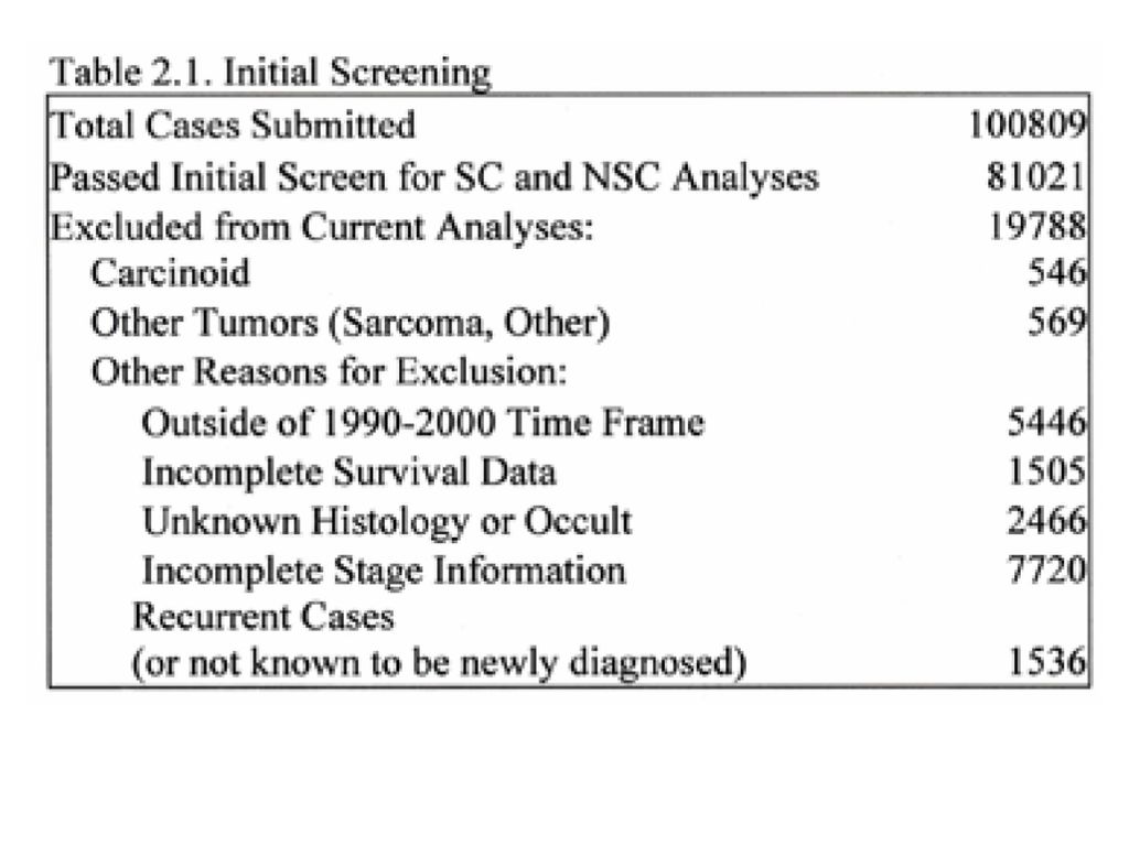
NSCLC : SCLC = 85% : 15% NSCLC = 30 : 30 : 40%
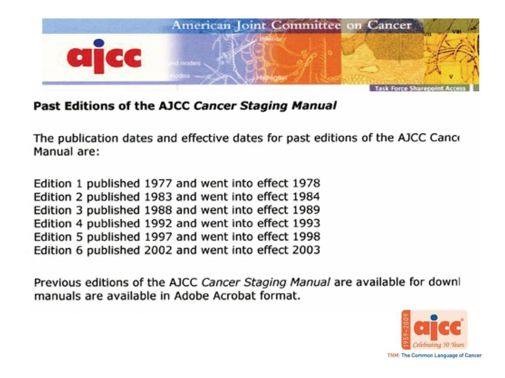
SCLC = 20 : 80% (10%이하에서 OP가능) 5, 6th edition 1997, 2002년: 5,319 cases of NSCLC 7th edition 2008년: 67,725 cases of NSCLC, 13,290 of SCLC
 5, 6th edition. 1997, 2002년: 5,319 cases of NSCLC. 7th edition. 2008년: 67,725 cases of NSCLC, 13,290 of SCLC.")
12
Stage TMN Descriptors IA T1 N0 M0 IB T2 N0 M0 IIA T1 N1 M0 IIB T2 N1 M0 T3 N0 M0 IIIA T3 N1 M0 T1–2–3 N2 M0 IIIB T4 N0–1–2 M0 T1–2–3–4 N3 M0 IV Any T any N M1

13
Tumor (T) Status Descriptor
Tumor <3 cm in greatest dimension, surrounded by lung or visceral pleura, without bronchoscopic evidence of invasion more proximal than lobar bronchus (i.e., not in main bronchus) T2 Tumor with any of following: >3 cm in greatest dimension; involves main bronchus, 2 cm distal to the carina; invades visceral pleura; associated with atelectasis or obstructive pneumonitis extending to hilum but does not involve entire lung T3 Tumor of any size that directly invades any of the following: chest wall (including superior sulcus tumors), diaphragm, mediastinal pleura, parietal pericardium; or tumor in main bronchus <2 cm distal to carina but without involvement of carina; or associated atelectasis or obstructive pneumonitis of entire lung T4 Tumor of any size that invades any of the following: mediastinum, heart, great vessels, trachea, esophagus, vertebral body, carina; or tumor with a malignant pleural or pericardial effusion, or with satellite tumor nodule(s) within the ipsilateral primary-tumor lobe of the lung
 T2. Tumor with any of following: >3 cm in greatest dimension; involves main bronchus, 2 cm distal to the carina; invades visceral pleura; associated with atelectasis or obstructive pneumonitis extending to hilum but does not involve entire lung. T3. Tumor of any size that directly invades any of the following: chest wall (including superior sulcus tumors), diaphragm, mediastinal pleura, parietal pericardium; or tumor in main bronchus <2 cm distal to carina but without involvement of carina; or associated atelectasis or obstructive pneumonitis of entire lung. T4. Tumor of any size that invades any of the following: mediastinum, heart, great vessels, trachea, esophagus, vertebral body, carina; or tumor with a malignant pleural or pericardial effusion, or with satellite tumor nodule(s) within the ipsilateral primary-tumor lobe of the lung.")
14
Lymph Node (N) Involvement Descriptor
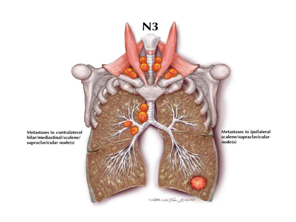
No regional lymph node metastasis N2 Metastasis to ipsilateral peribronchial and/or ipsilateral hilar lymph nodes, and intrapulmonary nodes involved by direct extension of the primary tumor N3 Metastasis to contralateral mediastinal, contralateral hilar, ipsilateral or contralateral scalene, or supraclavicular lymph node(s) Distant Metastasis (M) Descriptor M0 No distant metastasis M1 Distant metastasis presentb bSeparate metastatic pulmonary tumor nodule(s) in the ipsilateral non-primary tumor lobe(s) of the lung are classified as M1.
 Distant Metastasis (M) Descriptor M0. No distant metastasis. M1. Distant metastasis presentb bSeparate metastatic pulmonary tumor nodule(s) in the ipsilateral non-primary tumor lobe(s) of the lung are classified as M1.")
15
T1: T1a (≤2 cm in size) and T1b (>2-3 cm in size)
 and T1b (>2-3 cm in size)")
16
T2: T2a (>3-5 cm in size), T2b (>5-7 cm in size)
, T2b (>5-7 cm in size)")
17
T2 (>7 cm in size) ⇒ T3 T4 (Multiple nodules in the same lobe) ⇒ T3
 ⇒ T3 T4 (Multiple nodules in the same lobe) ⇒ T3")
18
M1: Multiple nodules in the same lung but a different lobe ⇒ T4

19
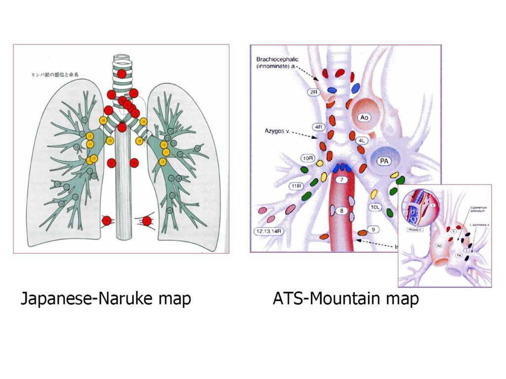
N1; peripheral zone (#12-14) and hilar-interlobar zone (#10, 11)
 and hilar-interlobar zone (#10, 11)")
20
N2; upper mediastinal zone (#1-4), aortopulmonary zone (#5, 6), subcarinal zone (#7), lower mediastinal zone (#8, 9)
, aortopulmonary zone (#5, 6), subcarinal zone (#7), lower mediastinal zone (#8, 9)")
22
Reclassify pleural dissemination as M1a
Subclassify M1 by additional nodule(s) in contralateral lung as M1a
 in contralateral lung as M1a.")
23
Subclassify M1 by distant metastases (outside the lung/pleura) as M1b
 as M1b")
25
The IASLC lymph node map

26
Stage TMN Descriptors IA T1a N0 M0 (≤2cm) T1b N0 M0 (2< to ≤3cm) IB
T2a N0 M0 (3< to ≤5cm) IIA T1a N1 M0 T1b N1 M0 T2a N1 M0 T2b N0 M0 (5< to ≤7cm) IIB T2b N1 M0 T3 N0 M0 (>7cm, same lobe) IIIA T4 N0 M0 (ipsilateral lung) T4 N1 M0 T3 N1 M0 T1-3 N2 M0 (T1a,b, T2a,b, T3) IIIB T4 N2 M0 T1–2–3–4 N3 M0 (T1a,b, T2a,b, T3, T4) IV Any T any N M1a (contralat. lung, pleural) Any T any N M1b Stage TMN Descriptors IA T1 N0 M0 IB T2 N0 M0 IIA T1 N1 M0 IIB T2 N1 M0 T3 N0 M0 IIIA T3 N1 M0 T1–2–3 N2 M0 IIIB T4 N0–1–2 M0 T1–2–3–4 N3 M0 IV Any T any N M1
 IIA. T1a N1 M0. T1b N1 M0. T2a N1 M0. T2b N0 M0 (5< to ≤7cm) IIB. T2b N1 M0. T3 N0 M0 (>7cm, same lobe) IIIA. T4 N0 M0 (ipsilateral lung) T4 N1 M0. T3 N1 M0. T1-3 N2 M0 (T1a,b, T2a,b, T3) IIIB. T4 N2 M0. T1–2–3–4 N3 M0 (T1a,b, T2a,b, T3, T4) IV. Any T any N M1a (contralat. lung, pleural) Any T any N M1b. Stage. TMN Descriptors. IA. T1 N0 M0. IB. T2 N0 M0. IIA. T1 N1 M0. IIB. T2 N1 M0. T3 N0 M0. IIIA. T3 N1 M0. T1–2–3 N2 M0. IIIB. T4 N0–1–2 M0. T1–2–3–4 N3 M0. IV. Any T any N M1.")
28
SCLC Stage: Limited disease(LD) vs Extensive disease(ED)
LD: confined to on hemithorax (although local extension: hilar, ipsilateral & contralateral mediastinal and supraclavicular nodes and ipsilateral pleural effusion regardless of cytology +/- if in the same radiation portal as the primary tumor and No extrathoracic metastases) IASCL (8088 SCLC) showed TMN staging is applicable to SCLC This edition should be applied to both NSCLC and SCLC
 IASCL (8088 SCLC) showed TMN staging is applicable to SCLC. This edition should be applied to both NSCLC and SCLC.")
29
Summary of Change Recommend for both NSCLC and SCLC and carcinoid tumors of lung T classification T1: T1a (≤2 cm in size), T1b (>2-3 cm in size) T2: T2a (>3-5 cm in size), T2b (>5-7 cm in size) T2 (>7 cm in size)→ T3 Multiple nodules in the same lobe: T4→ T3 Multiple nodules in the same lung but a different lobe: M1→ T4
, T1b (>2-3 cm in size) T2: T2a (>3-5 cm in size), T2b (>5-7 cm in size) T2 (>7 cm in size)→ T3. Multiple nodules in the same lobe: T4→ T3. Multiple nodules in the same lung but a different lobe: M1→ T4.")
30
N classification M classification M1: M1a, M1b
No changes However, New international LN map defining M classification M1: M1a, M1b Malignant pleural and pericardial effusions: T4→ M1a Separate tumor nodules in the contralateral lung : M1a M1b: distant metastases

31
감사합니다.

Similar presentations


 유명한 갯벌 ( 우리나라 ), 여러 갯벌 축제 갯벌이 만들어지는 조건 람사르 협약이란 ? 람사르 협약에 가입된 우리나라 생태지 밀물과 썰물 갯벌에.>")
 의 이해 흉부 기본 촬영 방사선 흉부 해부학 position of tubes and catheters.>")
 현장조사, 의식확인, 연락 현장은 안전한가 조사한다. 119 나 응급의료기관에 연락한다. 발바닥을 간지럽히거나 가볍게 꼬집어 본다. 0 ~ 4 분 4 ~ 6 분 6 ~ 10 분 10.>")


 1.>")
>")
>")

>")

, MELD 9) 로 F/U 중 2014.8월 HCC 3개 발견되어 TACE #1 차 시행후 f/u 중인 환자임. f/u MR 상 viable tumor (S5,>")
