Download presentation
Presentation is loading. Please wait.

1
분자생물학 실험 Western blot analysis

2
Introduction Western blot ( 때때로 ‘protein immunoblot’ 이라고 불림 ) 은 널리 알려진 분석 기술 중 하나로, 특정 단백질을 주어진 조직 샘플에서 인지하고자 할 때 이용된다. Gel electrophoresis(전기영동)으로 순수 단백질이나, 변성된 단백질을 삼차원 형태 그대로 크기 별로 분리된다. 크기 별로 분리 된 후에, 단백질들은 membrane(주로 nitrocellulose 나 PVDF 사용) 에 transfer한다. Membrane에 probe가 붙어있는 antibody들을 붙여줌으로써 찾고자하는 특정 단백질을 찾을 수 있다. Western blot 외에도 antibody를 사용하는 다른 기술들이 있는데, 조직이나 세포 내에서 단백질을 찾는 기술로 Immunostaining과 Enzyme linked immunosorbent assay(ELISA)가 있다. Western blot을 처음 만든 곳은 Stanford Univ.의 George Stark 교수님의 실험실이다. 그리고 ‘Western blot’이라는 명칭은, W. Neal Burnette과 Sushant Bhat에 의해 명칭 되었다. 이 명칭은 Edwin Southern에 의해 개발된, western과 비슷한 원리이지만 DNA를 인지하는 기술인 ‘Southern blot’을 따라서 붙여졌다. Western과 Southern과 비슷한 원리로 RNA를 분리하는 기술로는 Northern blot이 있다.
으로 순수 단백질이나, 변성된 단백질을 삼차원 형태 그대로 크기 별로 분리된다. 크기 별로 분리 된 후에, 단백질들은 membrane(주로 nitrocellulose 나 PVDF 사용) 에 transfer한다. Membrane에 probe가 붙어있는 antibody들을 붙여줌으로써 찾고자하는 특정 단백질을 찾을 수 있다. Western blot 외에도 antibody를 사용하는 다른 기술들이 있는데, 조직이나 세포 내에서 단백질을 찾는 기술로 Immunostaining과 Enzyme linked immunosorbent assay(ELISA)가 있다. Western blot을 처음 만든 곳은 Stanford Univ.의 George Stark 교수님의 실험실이다. 그리고 ‘Western blot’이라는 명칭은, W. Neal Burnette과 Sushant Bhat에 의해 명칭 되었다. 이 명칭은 Edwin Southern에 의해 개발된, western과 비슷한 원리이지만 DNA를 인지하는 기술인 ‘Southern blot’을 따라서 붙여졌다. Western과 Southern과 비슷한 원리로 RNA를 분리하는 기술로는 Northern blot이 있다.")
3
*분자생물학의 기본실험들 DNA를 검출할 수 있는 Southern blot RNA를 검출할 수 있는 Northern blot Protein을 검출할 수 있는 Western blot Western blot은 준비된 sample을 SDS-PAGE를 통해 단백질들을 분리하고 이 단백질들을 Membrane으로 옮기는 과정을 말하며 membrane으로 옮겨진 단백질은 항체를 이용하여 검출할 수 있다. 다음 그림은 transfer된 특정 단백질을 검출하는 원리이다.

4
<Western blot의 과정>
1. Tissue preparation

5
2. Gel electrophoresis

6
3. Transfer

7
4. Blocking & Antibody reaction

8
5. Detection Secondary antibody는 끝부분에 Horse radish peroxidase(HRP)가 결합되어 있는데 이를 화학적으로 처리하여 나오는 빛을 통해 band를 검출하게 된다. HRP는 luminol과 같은 substrate를 산화시키는데 산화된 luminol은 excited state가 되었다가 ground state 로 되면서 그 때의 에너지를 빛을 통해서 방출시킨다. 방출된 에너지는 film감광이나 빛으로 방출되는 에너지 파장을 검출함으로서 확인할 수 있다.
가 결합되어 있는데 이를 화학적으로 처리하여 나오는 빛을 통해 band를 검출하게 된다. HRP는 luminol과 같은 substrate를 산화시키는데 산화된 luminol은 excited state가 되었다가 ground state 로 되면서 그 때의 에너지를 빛을 통해서 방출시킨다. 방출된 에너지는 film감광이나 빛으로 방출되는 에너지 파장을 검출함으로서 확인할 수 있다.")
9
Purpose Western blot 의 원리를 이해하고 특정 단백질을 주어진 샘플에서 인지하여
단백질의 발현을 비교 분석 할 수 있다.

10
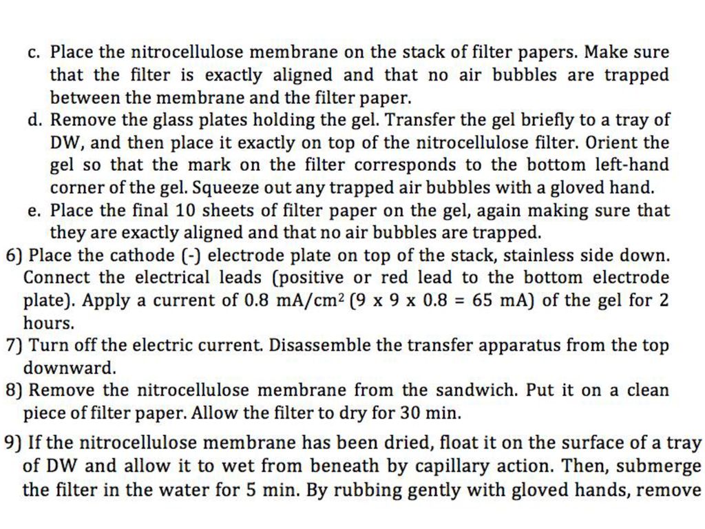
Method <Day 1> Running Samples
Pick 100 worms into 10 μl of Protease Inhibitor Cocktail in a 0.2-ml microfuge tube (and freeze at -70oC). Add 10 μl of 2X SDS sample buffer (make sure that 2-ME was pre-mixed) and boil for 10 min. & 4oC pause. Centrifuge briefly. Run 12 μl each of samples at 100 V through stack (top) gel, and at ~200 V (50 mA) while in separation (bottom) gel, until BPB dye reaches near the bottom end (but stop before it runs out of the gel).
. Add 10 μl of 2X SDS sample buffer (make sure that 2-ME was pre-mixed) and boil for 10 min. & 4oC pause. Centrifuge briefly. Run 12 μl each of samples at 100 V through stack (top) gel, and at ~200 V (50 mA) while in separation (bottom) gel, until BPB dye reaches near the bottom end. (but stop before it runs out of the gel).")
11
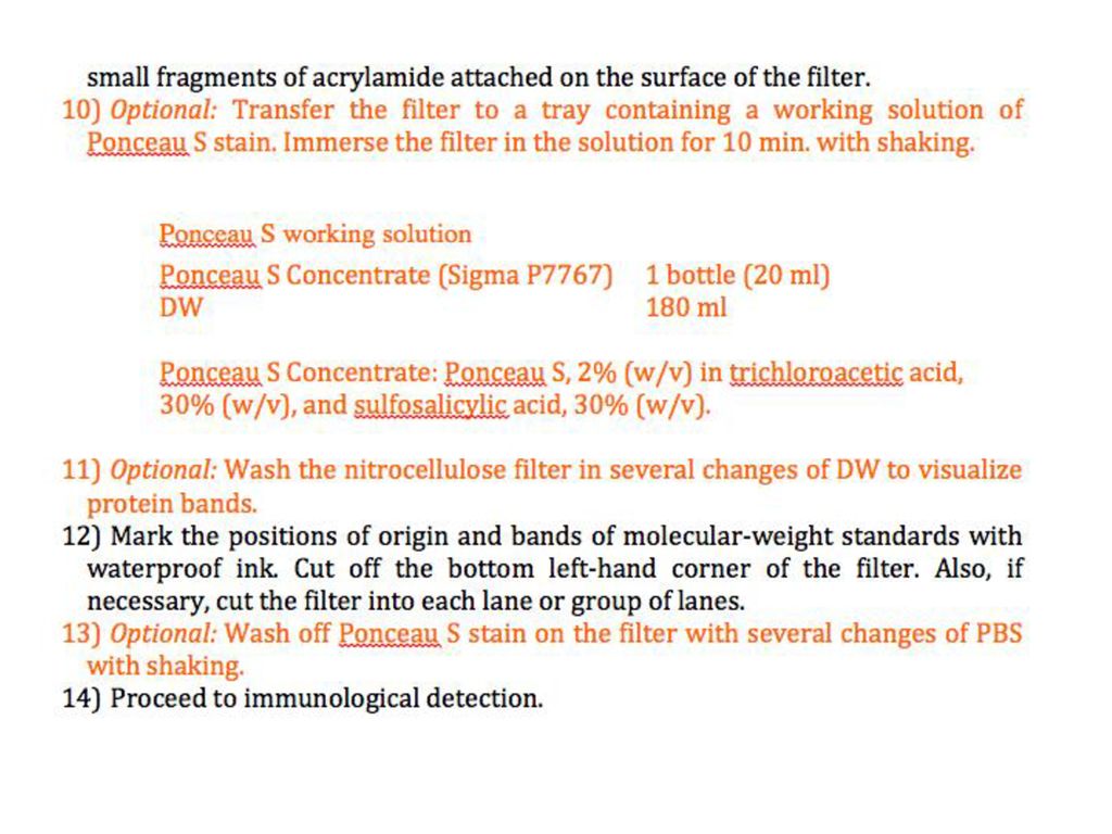
Western Blotting (Semi-Dry Transfer)
")
14
<Day 2> Immuno-detection

15
<Day 3>

16
Result Sample 1 Sample 2 Sample 3 Sample 4 Marker CED-4 Tubulin

17
CED-4 level is increased in pgl-1 and ife-1 mutants
N2(wild type) pgl-1(ct131) Ife-1(ok1978) ced-4(n1162) CED-4 Tubulin B CED-4 Topro3 d N2 CED-4 Topro3 SIR-2.1/tubulin intensity (% of control) N=100 d pgl-1 CED-4 Topro3 ife-1 d
 pgl-1(ct131) Ife-1(ok1978) ced-4(n1162) CED-4. Tubulin. B. CED-4. Topro3. d. N2. CED-4. Topro3. SIR-2.1/tubulin intensity. (% of control) N=100. d. pgl-1. CED-4. Topro3. ife-1. d.")
18
SIR-2.1/tubulin intensity
pgl-1 mutant showed higher level of SIR-2.1 cytoplasmic translocation than in N2 SIR-2.1 Topro-3 merged N2 d SIR-2.1 Topro-3 merged d pgl-1 Worms were harvested 48h after the L4 stage at 20℃. SIR-2.1 expression levels were not significantly changed in pgl-1 mutant. N=100 ife-1(ok1978) ced-4(n1162) sir-2.1(ok434) N2(wild type) pgl-1(ct131) SIR-2.1/tubulin intensity (% of control) SIR-2.1 Tubulin
 ced-4(n1162) sir-2.1(ok434) N2(wild type) pgl-1(ct131) SIR-2.1/tubulin intensity. (% of control) SIR-2.1. Tubulin.")
Similar presentations
>")
 은 여러 단백질의 혼합물로부터 찾고자 하는 단백질 에 대한 항체를 사용하여 항원 - 항체 반응을 일으킴 으로써 특정단백질을 찾아내는.>")
 5주차 Subcloning Ⅱ : Detection of Subcloning - Rapid Microscale Isolation of Plasmid from Transformed Cells 담당교수 : 하상준 교수님 담당조교 : 조소영.>")

 전기영동 (Electrophoresis) 전하를 띤 고분자 물질을 전기장을 띤 매질 ( 젤 ) 에서 이동 ∙ 분리 물질의 분리, 순도 ∙ 특성 분석 물질의 이동.>")


 1-2. The Sample list 1-1. The Method of Methylation.>")
>")


 미생물의 배양법 미생물이란 LB배지 미생물 배양 방법 고체배양 : 사면배양/ 천자배양 …>")
