Download presentation
Presentation is loading. Please wait.

1
KCP 723 삼성서울병원 전공의 하상윤

2
49세 여자 환자 피곤한 증상과 목의 혹 다수의 결절이 만져짐 초음파 검사: 검사실 소견
Rt: 2.0x1.0cm sized ill defined low echoic lesion, 0.4cm sized isoechoic nodule Lt: diffuse heterogeneous echogenicity, 1.3x1.3cm sized low echoic lesion multiple lymph node at bilateral level VI No suspicious lymph node on bilateral lateral neck 검사실 소견 CRP; 30.4mg/L (0.1~6.0) TSH receptor Ab; 1.24 IU/L (0~1.75) T3; ng/dL (71~161) Free T4; 2.0 ng/dL (0.8~1.7) TSH;<0.01 mIU/mL (0.86~4.69) Anti-TPO Ab; 19.5 IU/mL (5~13.6) Anti-thyroglobulin Ab; 18.4 IU/mL (10~124.2)
 TSH receptor Ab; 1.24 IU/L (0~1.75) T3; ng/dL (71~161) Free T4; 2.0 ng/dL (0.8~1.7) TSH;<0.01 mIU/mL (0.86~4.69) Anti-TPO Ab; 19.5 IU/mL (5~13.6) Anti-thyroglobulin Ab; 18.4 IU/mL (10~124.2)")
3
Cellular smear

4
염증세포가 background에 흩뿌려져 있고 핵이 주변 염증세포보다 큰 세포들이 모여 있는 것이 콜로이드와 함께 관찰되었습니다.크

5
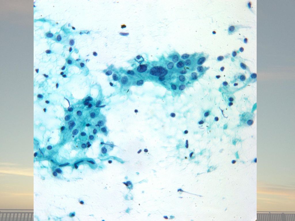
콜로이드와 follicular cell과 epitheliod histiocyte가 loose granuloma를 형성하고 있습니다.

6
follicular cell과 epitheliod histiocyte가 loose granuloma를 형성

7
Multinuleated giant cell
핵은 uniform하고 small round to oval sharp nuclear membrane finely granular chromain 하나정도의 micronucleoli Cytoplasm을 abundant granular. 올라와 있는 사진 끝

8
보내 주신 슬라이드 중 convenitonal smear 입니다.
저배율에서 역시 cellular smear를 볼 수 있으며 염증세포가 background에 관찰됩니다. 찍은 사진 Conventional 시작

9
Colloid를 먹고 있는 듯한, multinucleated giant cell
Colloid를 먹고 있는 듯한, multinucleated giant cell. dense한 cytoplasm을 가진 multinucleated giant cell 핵은 40개 정도3

10
Multinucleated giant cellconventional

11
Sheet of follicular cell

14
Scan conventional

15
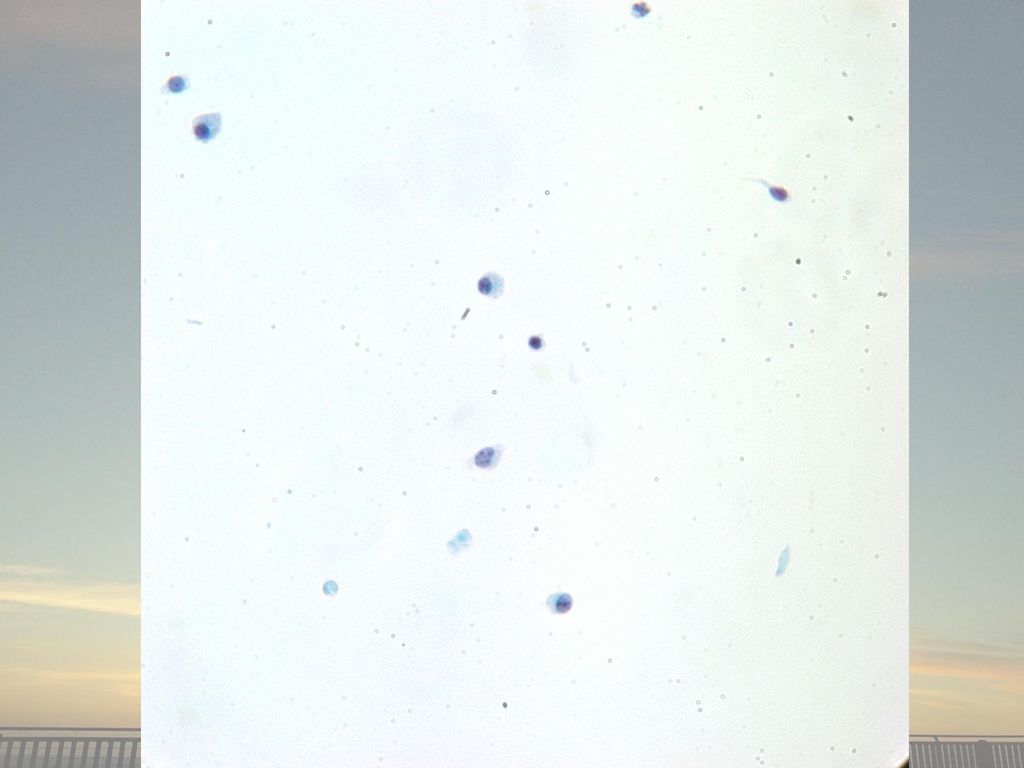
neutrophil

17
Thin prep 에서는 background의 염증세포는 많이 관찰되지 않았습니다.
Follicular cell들이 약간 핵이 커져 있고 nucleoli가 보이는 activated Follcular cell들이 sheet 형태로 관찰되었습니다.

18
여러 개의 multinucleated giant cell이 관찰되었습니다.

19
Histologic findings Cellular smear
Multinucleated giant cell around colloid Aggregation of epithelioid histiocytes Sheets of mildly atypical follicular cells No oncocytic cells A few colloid Some lymphocytes and histiocytes Few neutrophils and no plasma cells A few spindle stromal cells

20
Multinuclear giant cells in thyroid FNA
Nodular goiter or Thyroid neoplasm with cystic change (Foreign body histiocytic type) Variable in size Nuclei uniform and usually small in numbers Cytoplasm dirt, granular, ± hemosiderin Subacute thyroiditis (Multinucleated foreign body type) May be present in large numbers Enourmous in size with multiple, uniform nuclei in tens and hundreds Often seen in the vicinity of blobs of colloid, forming granulomas ± epithelioid cells Spindle shaped stromal cells Hashimoto’s thyroiditis (Multinuleated giant cell) Infrequent occurrence Nondescript morphology Fewer nuclei Associated Hashimoto’s features Palpation thyroiditis Acellular smear, rare chronic inflammatory cells Colloid follicular cells are usually absent
 Variable in size. Nuclei uniform and usually small in numbers. Cytoplasm dirt, granular, ± hemosiderin. Subacute thyroiditis. (Multinucleated foreign body type) May be present in large numbers. Enourmous in size with multiple, uniform nuclei in tens and hundreds. Often seen in the vicinity of blobs of colloid, forming granulomas ± epithelioid cells. Spindle shaped stromal cells. Hashimoto’s thyroiditis. (Multinuleated giant cell) Infrequent occurrence. Nondescript morphology. Fewer nuclei. Associated Hashimoto’s features. Palpation thyroiditis. Acellular smear, rare chronic inflammatory cells. Colloid follicular cells are usually absent.")
21
Multinuclear giant cells in thyroid FNA
Papillary carcinoma (Multinucleated giant cell) Variable in size Anaplastic carcinoma P/D metastatic carcinoma Bizarre nuclei with malignant criteria Infectious granulomatous lesions/ Sarcoidosis (Multinucleated foreign-body giant cells) Langhans-type giant cells and necrotic acellular debris characteristic in tuberculosis granulomas (AFB stain +) Associated with epitheliod cells and granulomas Inadvertent aspiration of thyroid or thyroid cartilage (Megakaryocytes) Multilobulated nuclei Smudgy chromatin Abundant dense cytoplasm Other hematopoietc cells in background Hodgkin’s lymphoma (RS cell) Varying sized, large to giant forms, mirror-image nuclei or multinucleated with prominent nucleoli
 Variable in size. Anaplastic carcinoma. P/D metastatic carcinoma. Bizarre nuclei with malignant criteria. Infectious granulomatous lesions/ Sarcoidosis. (Multinucleated foreign-body giant cells) Langhans-type giant cells and necrotic acellular debris characteristic in tuberculosis granulomas. (AFB stain +) Associated with epitheliod cells and granulomas. Inadvertent aspiration of thyroid or thyroid cartilage. (Megakaryocytes) Multilobulated nuclei. Smudgy chromatin. Abundant dense cytoplasm. Other hematopoietc cells in background. Hodgkin’s lymphoma. (RS cell) Varying sized, large to giant forms, mirror-image nuclei or multinucleated with prominent nucleoli.")
22
conventional

23
Hashimoto’s VS Subacute
Abundant mixed lymphocyte and plasma cells Germinal center cells Lymphohistiocytic aggregates Hurthle cells and variable nuclear atypia Infrequent multinucleated giant cell / absent granulomas Little colloid Many multinucleated giant cell (numerous nuclei), often in the vicinity of blobs of colloid Mixed inflmmatory cells including epitheliod histiocytes and lymphocytes Hypocellular smear: scant follicular cell Degenerative changes in follicular cells, cell debris and colloid
, often in the vicinity of blobs of colloid. Mixed inflmmatory cells including epitheliod histiocytes and lymphocytes. Hypocellular smear: scant follicular cell. Degenerative changes in follicular cells, cell debris and colloid.")
24
Hashimoto’s VS Subacute
Pain over the thyroidal bed was recorded in 100% of subacute thyroiditis. Typical cases of Hashimoto’s disease do not present pain over the thyroidal bed. Cytologic findings of the final stages of the two condition may overlap: very small numbers of lymphocytes and oncocytic cells Fine-needle aspiration of subacute granulomatous thyroiditis (De Quervain's thyroiditis) : a clinico-cytologic review of 36 cases. Diagn Cytopathol Mar;16(3):
 : a clinico-cytologic review of 36 cases. Diagn Cytopathol Mar;16(3):")
25
Hashimoto’s disease Subacute thyroiditis
49세 여자 환자 o 피곤한 증상 Thyroid nodules Neck LN enlargement ? CRP; 30.4mg/L (0.1~6.0) T3; ng/dL (71~161) Free T4; 2.0 ng/dL (0.8~1.7) TSH;<0.01 mcIU/mL (0.86~4.69) Anti-TPO Ab; 19.5 IU/mL (5~13.6) Anti-thyroglobulin Ab; 18.4 IU/mL (10~124.2)
 T3; ng/dL (71~161) Free T4; 2.0 ng/dL (0.8~1.7) TSH;<0.01 mcIU/mL (0.86~4.69) Anti-TPO Ab; 19.5 IU/mL (5~13.6) Anti-thyroglobulin Ab; 18.4 IU/mL (10~124.2)")
26
검사실 소견의 해석 The prevalence of elevated serum C-reactive protein levels in inflammatory and noninflammatory thyroid disease. Thyroid Jul;13(7):643-8.
:")
27
검사실 소견의 해석 Salivary C-Reactive Protein in Hashimoto’s Thyroiditis and Subacute Thyroiditis International Journal of Inflammation Volume 2010, Article ID , 5 pages

28
검사실 소견의 해석 Acute-phase reactants in Hashimoto thyroiditis
Int Immunopharmacol Dec 20;8(13-14):1863-5
:")
29
검사실 소견의 해석 Anti-TPO Ab were detected
TSH TT3 FT4 TPO-Ab CRP Subacute <0.005 mU/L (1.1±0.7) 2.2±0.36 ng/ml (1.09±0.25) 21.6±6.8 pg/mL (10.0±1.5) 0% 33.4±26.4 mg/L (2.6±4.0) Hashimoto 1.9±1.6 0.48±0.25 9.9±2.7 100% 2.8±4.1 KCP 723 0.01 mIU/mL ( ) 160.5 ng/dL (71-161) 2.0 ( ) 19.5 IU/mL (5-13.6) 30.4 ( ) Anti-TPO Ab were detected in 10 of 119 (8.4%: range, U/mL) in normal controls. Antithyroid peroxidase autoantibodies in thyroid diseases. J Clin Endocrinol Metab Sep;71(3):661-9.
 2.2±0.36. ng/ml. (1.09±0.25) 21.6±6.8. pg/mL. (10.0±1.5) 0% 33.4±26.4. mg/L. (2.6±4.0) Hashimoto. 1.9± ± ± % 2.8±4.1. KCP mIU/mL. ( ) ng/dL. (71-161) 2.0. ( ) 19.5 IU/mL. (5-13.6) ( ) Anti-TPO Ab were detected. in 10 of 119 (8.4%: range, U/mL) in normal controls. Antithyroid peroxidase autoantibodies in thyroid diseases. J Clin Endocrinol Metab Sep;71(3):")
30
DIAGNOSIS Some multinucleated giant cells, epthelioid histiocytes aggregation, and mildly aypical follicular cells in the background of lymphocytes and a few colloid (s.n) Note) 임상적으로 pain이 있다면 subacute thyroiditis의 가능성이 높습니다.
 Note) 임상적으로 pain이 있다면 subacute thyroiditis의 가능성이 높습니다.")
31
경청해 주셔서 감사합니다.

Similar presentations


. CC: Low abd distension for 1 M CC: Low abd distension for 1 M (Pap: N these day, CA125: 15) (Pap: N these day, CA125: 15) Wt loss.>")

 제출자 발표 2012.02.01 서울성모병원 병리과 전공의 이영섭.>")
 2) 3) 4) 2. 1) 2) 3. 1) 2) 3) * 혈액의 생성과 파괴 혈구생성부위 -- 1. 2. 3. * 조혈자극인자 –- 1. 2. 3. * 적혈구의 분화와 성숙과정 – Pluripotent stem cell →>")


>")


>")

![MGR review 류마티스 내과 R4 최인아. [ 목차 ] Overlap syndrome (vs UCTD, MCTD) Polymyositis(PM) PM with Interstitial lung disease.](/42/11353284/big_thumb.jpg "MGR review 류마티스 내과 R4 최인아. [ 목차 ] Overlap syndrome (vs UCTD, MCTD) Polymyositis(PM) PM with Interstitial lung disease.>")

 비만률순위 : 29 위 (2005 OECD)>")

