Download presentation
Presentation is loading. Please wait.

1
GLAUCOMA CONFERENCE 안압 측정, 전방각 검사 Ap.박명희/R3 김성일

2
정상 안압 Distribution 10~21mmHg(mean IOP 15.5±2.57mmHg)
skewed distribution

3
안압에 영향을 미치는 요소 Factors exerting long-term influence on intraocular pressure Genetics Age in general incerease with age reduced facility of aquous outflow, uveoscleral outflow reduced aquous production Gender equal between 20~40 yrs increase in women coincide with menopause Refractive error high myopia have high incidence of chronic open angle glaucoma Ethnicity

4
안압에 영향을 미치는 요소 Factors exerting short-term influence on intraocular pressure diurnal variation 주기적으로 변하며 정상 변동치는 3~6mmHg 변동폭이 10mmHg이상이면 병적으로 본다 postural variation 앉은 자세에서 바로 누우면 0.3~6.0mmHg 상승 개방각녹내장이나 정상안압녹내장 환자에서 변동폭이 크다 exertional influence 격렬하거나 장시간 운동은 안압을 하강시킴

5
안압에 영향을 미치는 요소 Factors exerting short-term influence on intraocular pressure lid and eye movement 눈 깜박임은 안압을 10mmHg 정도 상승시킴 심하게 감으면 90mmHg까지도 상승, 지속적으로 감으면 하강 벨마비에서는 하강 intraocular conditions anterior uveitis rhegmatous retinal detachment systemic conditions 수축기 혈압이 상승하면 안압도 상승함 environmental conditions general anesthesia 일반적으로 안압 하강 발생(예외-ketamine) Depolarizing muscle relaxants(succinylcholine)은 일시적 안압 상승 food and drugs
 Depolarizing muscle relaxants(succinylcholine)은 일시적 안압 상승. food and drugs.")
6
안압의 측정 안압의 측정방법 Indentation tonometer Applanation tonometer
표준 무게에 따른 각막의 합입을 측정 Schoitz Applanation tonometer 각막을 일정 면적 편평하게 하는데 필요한 힘을 통해 계산 Goldman tonometer Perkins tonometer Non-contact tonometer(pneumatic tonometer) 공기를 분사한 후 각막이 편평해지는 데까지 걸린 시간 으로 계산
 공기를 분사한 후 각막이 편평해지는 데까지 걸린 시간 으로 계산.")
7
Goldman applanation tonometer
기본개념 Imbert-Fick law 각막을 일정 면적으로 편평하게 만드는데 필요한 힘을 통 해 계산 (W=Pt x A) 완전 구형, 두께가 무한대로 얇고 건조하다는 가정 압평면의 직경이 3.06mm2일때 tear film의 표면장력과 각막의 저항력 상쇄 오차의 요인 반윤상의 넓이가 넓으면 높게 측정 안구를 누르거나 감으려고 하면 높게 측정 각막의 두께가 얇으면 낮게 측정 각막 부종시 낮게 측정 각막 굴절력이 높아지면 높게 측정(4디옵터당 1mmHg) 직난시에서 낮게 측정
 완전 구형, 두께가 무한대로 얇고 건조하다는 가정. 압평면의 직경이 3.06mm2일때 tear film의 표면장력과 각막의 저항력 상쇄. 오차의 요인. 반윤상의 넓이가 넓으면 높게 측정. 안구를 누르거나 감으려고 하면 높게 측정. 각막의 두께가 얇으면 낮게 측정. 각막 부종시 낮게 측정. 각막 굴절력이 높아지면 높게 측정(4디옵터당 1mmHg) 직난시에서 낮게 측정.")
8
전방각경 검사

9
전방각의 구조

10
전방각경의 원리 Critical angle

11
Direct gonioscope Koeppe lens Gonioscope : 15 to 20 x magnification
Light source: usually a separate handheld unit Patient in a supine position Bridge balanced salt solution : methylcellulose or patient’s own tear film Scan the anterior chamber angle by shifting patient’s position
12
Indirect gonioscope Viewed indirectly through mirror 180 degrees
from the quadrant being viewed Goldmann single mirror lens Goldmann three mirror lens two mirrors for examination of fundus one mirror tilted 59 for ant.chamber angle Zeiss four mirror lens four mirror are tilted 64 degrees eliminate the need to rotate

13
Goldmann three-mirror

14
Compressive Gonioscopy
To distinguish between appositional and synechial closure of the angle

15
전방각경의 장단점 비교

16
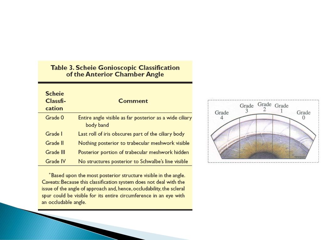
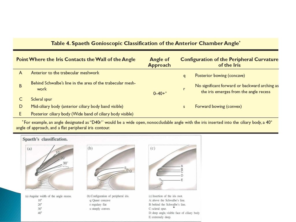
Grading Systems

21
시신경유두검사

22
Normal optic nerve head
Definition Distal portion of optic nerve, directly susceptible to elevated IOP Anatomy Size(V x H): 1.88 x 1.77 mm(vertically oval) Disc area: 0.68 ~ 4.42 mm2 (2.42 mm2) Vasculature Arterial supply Post. Ciliary artery central retinal artery의 arteriolar branch Venous drainage Central retinal vein choroid
: 1.88 x 1.77 mm(vertically oval) Disc area: 0.68 ~ 4.42 mm2 (2.42 mm2) Vasculature. Arterial supply. Post. Ciliary artery. central retinal artery의 arteriolar branch. Venous drainage. Central retinal vein. choroid.")
23
Morphology of the Normal Optic Nerve and Head
General feature - Vertically oval - Cup - central porion of depression - horizontally oval - Pallor - absence of axon - Neural rim - location of bulk of the axon - normally orange-red color because of the associated capillaries - ISNT rule - larger disc have larger neural rim area

24
Morphology of the Normal Optic Nerve and Head
Cup/Disc ratio (C/D) -only indirect measure of the amount of neural tissue : may be misleading larger diameter of nerve head – thinner neural rim width and larger cup size, despite a stable number of axon. : attention to appearance of neural rim.
 -only indirect measure of the amount of neural tissue. : may be misleading. larger diameter of nerve head – thinner neural rim width and larger cup size, despite a stable number of axon. : attention to appearance of neural rim.")
25
Morphology of the Normal Optic Nerve and Head
Physiologic peripapillary retina Retinal nerve fiber layer - 정상적으로 striation이 관찰됨(nerve fiber의 light reflex) - Nerve bundle이 criical thickness 이상이어야 보임 - Posterior pole과 peripapillary region 에서 잘 관찰, - vertical pole에서 temporal로 향하는 bundle이 가장 잘 보 임 Peripapillary pigmentary variations cup neuroretinal rim scleral lip thin, even, white rim zone beta chorioscleral crescent (RPE retraction) zone alpha malposition of the embryonic fold with a double layer or irregularity of RPE
 - Nerve bundle이 criical thickness 이상이어야 보임. - Posterior pole과 peripapillary region 에서 잘 관찰, - vertical pole에서 temporal로 향하는 bundle이 가장 잘 보 임. Peripapillary pigmentary variations. cup. neuroretinal rim. scleral lip thin, even, white rim. zone beta chorioscleral crescent (RPE retraction) zone alpha malposition of the embryonic fold with a double layer or irregularity of RPE.")
26
Morphology of Glaucomatous Optic Atrophy
Disc pattern Focal atrophy Concentric atrophy Deepening of the cup Pallor/cup discrepancy Vascular signs Optic disc hemorrhage Tortuosity of retinal vessels Cilioretinal arteries Localtion of retinal vessels Peripapillary changes Nerve fiber bundle defects Peripapillary pigmentary disturbance

27
Focal atrophy Selective loss of neural rim Focal notching
primarily in the inferotemporal region enlargement of cup in a vertical or oblique direction Vertical poles temporal nasal Focal notching small, discrete defect (usually inferotemporal quadrant) Sharpened rim local thinning of neural rim tissue reaches the disc margin Bayoneting
 Sharpened rim. local thinning of neural rim tissue reaches the disc margin. Bayoneting.")
28
Concentric atrophy generalized expansion of the cup retention of its “round” appearance early glaucomatous damage의 가장 흔한 form compare in the fellow eye for symmetry 혹은 serial photograph를 통하여 physiologic cup을 감별

29
Pallor/cup discrepancy
Deepening of the cup Overpass cupping Initially bridge the deepened cup, later collapse into it Laminar dot sign Grey fenestra of the lamina Dot-like or striated Pallor/cup discrepancy In the early stage, Enlargement of cup progress ahead of pallor Vessel의 kinking이나 입체적인 검사법으로 cupping 을 파악해야 함. Saucerization early glaucomatous change의 일종. diffuse, shallow cupping extend to the disc margins with retention of a central pale cup early sign of glaucoma

30
Advanced glaucomatous cupping total cupping , white disc
eventual loss of all neural tissue Bean-pot cupping

31
Vascular Signs of Glaucomatous Optic Atrophy
Optic disc hemorrhages splinter hemorrhage More common in NTG than POAG or glaucoma suspect tend to come & go Usually near the margin of the optic nerve head Most common in inferior quadrant More common in early to middle stage of damage

32
Tortuosity of retinal vessel
on the disc advanced glaucomatous optic atrophy에서 볼 수 있음. Chronic CRVO에 대한 response로 collateral vessel이 loop를 형성한 것으로 보여짐.

33
Glaucomatous change
34
Cilioretinal arteries
glaucomatous eye with one or two temporal cilioretinal arteries cilioretinal artery가 없는 group에 비해서 중심시야가 보다 잘 보존될 수 있다는 보고가 있음.

35
Location of retinal vessel
in relation to the cup. Some diagnostic value Overpass cupping Nasal displacement of vessels function of cup size Baring of the circumlinear vessel 시야결손과 유의한 관련이 있다. Retinal vessel beyond the disc margin may also undergo glaucomatous change (ex. general arterial narrowing)
")
36
Nerve fiber bundle defect
loss of nerve bundle visible defect in the retinal nerve fiber layer dark strip or wedge shape defects visual field change, neural rim area, flourescein filling defect 와 밀접한 연관 Normal striation Slit-like defect

37
Peripapillary pigmentary disturbance
also seen in other condition myopia, aging Peripapillary atrophy both zone beta and zone alpha more frequently and larger in glaucomatous eye than in normal progressively enlarge with glaucomatous change Absence of peripapillary atrophy lower the risk of glaucomatous damage among patients ocular hypertension

Similar presentations