Download presentation
Presentation is loading. Please wait.

1
응급실에서 접하는 의학적 영상 Chest Radiograph (Plain) Brain CT 건국대학교 충주병원 영상의학과
응급실에서 접하는 의학적 영상 Chest Radiograph (Plain) Brain CT 건국대학교 충주병원 영상의학과 최 영 칠
 Brain CT. 건국대학교 충주병원 영상의학과. 최 영 칠.")
2
응급실에서 접하는 의학적 영상 I - Chest Radiograph

3
조직의 X선 감약도 . (이 순서대로 크다 – 사진상 희다) 1. Bone density; 골 2. Water density;
근육, 혈액, 삼출액, 흉수등 3. Fat density; 지방 4. Air density; 공기 .

4
흉부X선사진에서 볼 것 1. 기관지와 폐혈관을 포함하는 폐실질 종격동과 심장 및 대혈관 횡경막과 흉벽 및 흉곽의 골격

5
폐실질; 폐엽의 구분

6
정상 Chest PA and Lateral

8
Alveolar Dz. Airway Dz. Interstitial Dz. Vascular Dz. Neoplastic Dz
* Alveolar Dz * Airway Dz * Interstitial Dz * Vascular Dz * Neoplastic Dz * Mediastinal Dz * Pleural, Diaph. and Th. Wall Dz * Congenital Dz

9
Alveolar vs Interstitial lesion

10
Silhouette sign

11
Hematogeneous vs Lymphangitic Metastasis

12
Diffuse nodular lesion

13
Atelectasis

14
Pneumonia

15
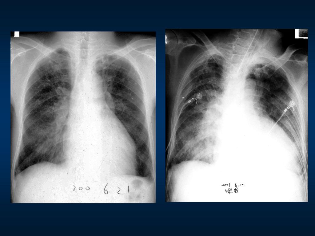
Tuberculosis

16
Bronchiectasis

17
Lung cancer

18
Emphysema

19
Trauma

20
Diaphragmatic Hernia

21
Effusion

22
.

23
응급실에서 접하는 의학적 영상 II - Brain CT

24
정상 brain CT

25
정상 brain CT

26
* Stroke * Trauma * Tumor * Infection * Congenital Anomaly

27
I. Stroke; loss of a neurologic function secondary to parenchymal
A new, often acute, loss of a neurologic function secondary to parenchymal ischemia or hemorrhage.

28
A. Cerebral infarction Large vessel occlusions
Small vessel infarctions Cardiac emboli Blood disorders Vasculitis...

29
Vascular Territories

30
Hyperacute infarction (<12 hours, or within first few hours)
Normal (50% to 60%) Hyperdense artery sign Obscuration of lentiform nucleus
 Hyperdense artery sign. Obscuration of. lentiform nucleus.")
31
Acute Infarction (12 - 24 hours) Low density in basal ganglia
Loss of gray-white matter differentiation (loss of insular ribbon, obscuration of cortex-white matter border) sulcal effacement
 sulcal effacement.")
32
Acute Infarction (1 - 3 days) (4 - 7 days) Increasing mass effect
Wedge-shaped low density involves both gray and white matter Hemorrhagic transformation may occur (4 - 7 days) Gyral enhancement Mass effect, edema persists
 Gyral enhancement. Mass effect, edema persists.")
33
B. Primary intracranial hemorrhage
Very common Hypertension Aneurysm Vascular malformation Prematurity

34
Location of Hypertensive Hemorrhage
Putamen/external capsule 60% to 65% Thalamus 15% to 25% Pons 5% to 10% Cerebellum 2% to 5% Subcortical white matter 1% to 2%

35
Imaging of ICH Acute Subacute Chronic

36
C. Nontraumatic SAH Aneurysm Vascular malformation

37
II. Trauma Traumatic Craniocerebral Lesions Skull & Scalp lesions
Extracerebral hemorrhage EDH SDH SAH Intraaxial lesions Diffuse axonal injury (shearing injury) Cortical contusion Deep cerebral gray matter and brainstem injury Intraventricular/choroid plexus hemorrhage
 Cortical contusion. Deep cerebral gray matter and brainstem injury. Intraventricular/choroid plexus hemorrhage.")
38
Skull fracture, Scalp hematoma/laceration

39
Cortical contusion

40
Epidural hematoma

41
Subdural hematoma Acute Subacute Chronic

42
Subarachnoid hemorrhage

43
Diffuse axonal injury (shearing injury)
")
Similar presentations


 의 이해 흉부 기본 촬영 방사선 흉부 해부학 position of tubes and catheters.>")


 주요 내용 소개>")


 2010. 03. 12. 흉부외과 최 주 원.>")


