Download presentation
Presentation is loading. Please wait.

1
시신경 검사시 녹내장과 감별해야할 시신경 질환들
가톨릭대학교 의과대학 안과 및 시과학 교실 R4 장동진 / Pf. 문정일

2
Contents Approach to ONH Cases to DDx from Glaucoma
시신경 유두를 관찰하는 것이 녹내장 진단의 시작이며, 이는 안압과 함께 건강검진에서 보는 두가지 검사임. 이 발표의 목적은 진료실에서 시신경 유두 검사를 할때 어떻게 접근할 것이며, 이 모양이 정상적인지 아닌지, 아니라면 녹내장과 어떻게 감별을 할 수 있을지에 대하여 알아보고자 하는 것임.

3
Before We Start

4
Approaches OD OS CDR(V/H) 0.6/0.6 LDS + PPA Pallor - Hemorrhage
We record like below OD OS CDR(V/H) 0.6/0.6 LDS + PPA Pallor - Hemorrhage Nasalization Notching Bayoneting RNFL defect 기본적으로 환자의 나이, 성별, 가족력 등을 확인 한 후에 안과적 검사를 하게되면, 이는 우리가 시신경을 볼때 관찰하는 항목들이며 양안을 같이 비교하는 것도 중요, rim defect, ISNT rule을 따르는지 볼것. re 우리가 챠팅할때 보는 것이지만 닫순히 이 항목에만 집중해서는 전반적인 그림을 보기가 어려울 수 있다.
 0.6/0.6. LDS. + PPA. Pallor. - Hemorrhage. Nasalization. Notching. Bayoneting. RNFL defect. 기본적으로 환자의 나이, 성별, 가족력 등을 확인 한 후에 안과적 검사를 하게되면, 이는 우리가 시신경을 볼때 관찰하는 항목들이며 양안을 같이 비교하는 것도 중요, rim defect, ISNT rule을 따르는지 볼것. re. 우리가 챠팅할때 보는 것이지만 닫순히 이 항목에만 집중해서는 전반적인 그림을 보기가 어려울 수 있다.")
5
General Features ONH size? Vertical oval shape? Neural Rim ISNT rule Gray crescent Peripapillary retina RNFL Peripapillary pigmentary variations Cup size - CDR(V/H) shape - horizontally oval, V>H?
 shape - horizontally oval, V>H")
6
Let’s Dig in! Keep in mind CDR(V/H) concentric atrophy 양안 비교 LDS*
deepening of the cup PPA zone alpha and zone beta 양안비교 Pallor Pallor-cup discrepancy, Saucerization Hemorrhage Splinter hm. may be the first sign Nasalization Watch the vsls Notching focal rim notching 찾아라! 초기에 나타남 Bayoneting Loss of neural rim RNFL defect dark stripes or wedge-shape *LDS: laminar dot sign

8
Gray crescent Physiologic Large Cup
9
Physiologic Large Cup
10
Physiologic Large Cup Rather a exclusion diagnosis! Do other exams!
선천적 양안 large optic nerve head with cup↑ 정상 rim area, 시야, 안압 Axon이 large surface disc로 퍼져 disc surface와 neuroretinal rim pallor 대개 optic disc cup은 round하거나 horizontally oval-shaped 후천적 어린이 orbital glioma: 이전 정상크기 optic disc 점점 ↑ 경우 녹내장과의 감별 rim pallor(+), notch(-), nerve fiber layer defects(-)
, notch(-), nerve fiber layer defects(-)")
11
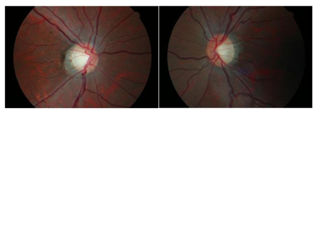
Glaucoma? VF- normal IOP - normal nearly total cupping pallor
RNFL defect? CDR(V/H)LDSPPAPallorHemorrhageNasalizationNotchingBayonetingRNFL defect Glaucoma? VF- normal IOP - normal
12
Optic Disc Coloboma Pathogenesis : defective closure of the embryonic fetal fissure Sporadically or AD 유전(renal-coloboma syndrome : PAX2 gene mutation) Bowl-shaped excavation, decentered (아래쪽에 발생) Unilateral or bilateral Iris coloboma, retinochoroidal coloboma가 함께 관찰될 수 있음. Visual field changes Arcuate scotoma Generally not progressive Progressive : autosomal dominant type
 Bowl-shaped excavation, decentered (아래쪽에 발생) Unilateral or bilateral. Iris coloboma, retinochoroidal coloboma가 함께 관찰될 수 있음. Visual field changes. Arcuate scotoma. Generally not progressive. Progressive : autosomal dominant type.")
13
Optic Disc Coloboma
15
Optic Disc Pit Atypical coloboma Unilateral: > 80%
Larger disc on the involved side Association with serous retinal detachment: 40 % Localized pale and deep depression, near the temporal or inferotemporal side Visual field changes Paracentral or arcuate scotoma in 50% of cases Enlarged blind spot

18
Superior segment optic hypoplasia (Topless Optic Disc syndrome)
Congenital anomaly Usually bilateral Associated with diabetic mother: 8.8% of prevalence Findings Relative superior entrance of the central retinal artery Thinning of the superior peripapillary nerve fiber layer Superior peripapillary scleral halo Pallor of the superior disc Visual field changes Sector like defect

20
Tilted Disc Syndrome Abnormal closure of the embryonic fissure
Tilted to the inferior nasal side Inferior or inferonasal crescent Associated with myopia, astigmatism Tilted its horizontal axis Inferior chorioretinal hypoplasia Visual field changes Superotemporal field loss Incomplete bitemporal depression: not respect vertical midline Enlarged blind spot Non progressive

22
Autosomal Dominant Optic Neuropathy
OPA1 gene on chromasome 3→dynamin-related protein(a family of GTPase) :NTG의 marker로도 여겨 짐 Pathology : primary degeneration of retinal ganglion cells with ascending optic atrophy Family history History of gradual, bilateral loss of vision Dyschromatopsia Central scotoma Pallor of the remnant rim
 :NTG의 marker로도 여겨 짐. Pathology : primary degeneration of retinal ganglion cells with ascending optic atrophy. Family history. History of gradual, bilateral loss of vision. Dyschromatopsia. Central scotoma. Pallor of the remnant rim.")
23
어떤 나이에서라도 (특히 10-20세) 양안의 설명할 수 없는 시신경병증 서서히 진행할 때 부모나 형제가 증상이 없더라도 안과 검사 시신경위축이나 경미한 변화
 양안의 설명할 수 없는 시신경병증 서서히 진행할 때 부모나 형제가 증상이 없더라도 안과 검사 시신경위축이나 경미한 변화")
Similar presentations


 광양 매화마을 (40m) 사천 와룡산 철쭉 (1h) 사천 한려수도 (1h) 순천 순천만 갈대밭 (1h) 순천 삼보사찰 송광사 (1h20m) 구례 산수유마을 (1h30m) 산청 웅석계곡 (1h30m) 거제 외도 (1h50m)>")

 제출자 : 9741232 조한진 9741203 이영수 0071250 이호진 동아일보에 사용된 그래프의 오남용 사례 분석.>")

 주요 내용 소개>")


 Brain CT 건국대학교 충주병원 영상의학과>")


